Nephrology

13 Nephrology


The manifestations of renal and genitourinary disorders range from readily apparent gross structural abnormalities to subtle abnormalities of the urinary sediment. In this chapter, examples of physical findings, as well as characteristic urinary findings and radiographs, are used to demonstrate the broad spectrum of these disorders in the pediatric population.


Essentials of Medical History and Physical Examination


The medical history and physical examination often provide clues implicating a renal or genitourinary disorder. Congenital but often nonheritable genitourinary disorders are diagnosed with increasing frequency by high-resolution ultrasonography during the second and third trimesters and may include obstructive disorders, such as posterior urethral valves, multicystic dysplasia, polycystic kidney disease, prune-belly syndrome, and renal agenesis. Oligohydramnios and fetal compression signs reflect reduced urine production associated with some of these disorders and may result in early postnatal death as a result of associated pulmonary underdevelopment. Unilateral and, less frequently, bilateral cystic dysplasia is the most common cause of abdominal mass in newborns. A large placenta may be a telltale sign of congenital nephrotic syndrome of the Finnish type, in which severe proteinuria precedes birth. Failure to urinate during the first 24 hours of life should prompt evaluation for obstruction of the kidneys, ureters, or bladder. Urinary tract infection (UTI) should be a consideration in all febrile infants, particularly during the first month, even in the presence of documented sepsis, meningitis, or other sources of infection. Urinary tract anomalies, including vesicoureteral reflux and megaureters distending the abdomen, are common in infants and young children with well-documented UTI. Children with true polyuria or polydipsia, rather than urinary frequency, may have a renal concentrating defect, such as nephrogenic diabetes insipidus, or salt-losing nephropathy, such as nephronophthisis.


Failure to thrive, lethargy, or irritability and recurrent emesis are common manifestations of renal disease in infants and may be associated with metabolic acidosis and other electrolyte disturbances. On occasion, the renal disorder is discovered because of deliberate studies obtained after discovering dysmorphic features, imperforate anus, vertebral abnormalities, fetal alcohol syndrome, or other disorders that have a renal component.


Family pedigrees may facilitate the diagnosis of congenital or heritable disorders such as cystinuria, cystinosis, oxaluria, and polycystic kidney disease and thereby lead to a variety of preventive measures before children become symptomatic. Hypertension in infants without aortic coarctation is most often a result of a renovascular disorder, such as renal venous thrombosis, arterial thrombosis (caused by embolism in patients with ventricular septal defect or patent ductus arteriosus, or a result of umbilical artery catheterization), or renal artery stenosis. Asphyxia at birth or severe dehydration, sepsis, or shock may lead to acute tubular necrosis and oliguric acute kidney injury.


Hypertension in the older child may cause headache, dizziness, recurrent emesis, epistaxis, or visual disturbances. In severe cases secondary congestive heart failure may occur, particularly if there is a history of oliguria, impaired renal function, or glomerulonephritis. Renal and renal vascular disorders account for most cases of hypertension, especially in school-age children and younger. Primary hypertension has become the most common cause in adolescents, often in association with obesity or metabolic syndrome. The presence of café-au-lait spots, neurofibromas, fibrous-angiomatous lesions of the skin, thyroid enlargement, abnormal pulses, or bruits over the renal arteries or major vessels may point to a specific diagnosis.


Gross or microscopic hematuria is the most common reason children are referred to outpatient pediatric nephrology clinics. The medical history is critical to pinpointing the correct cause of hematuria because it facilitates the elimination of a large number of possibilities. These possibilities include complications in the neonatal period necessitating umbilical artery line placement that may result in renal or aortic occlusive disease, bronchopulmonary dysplasia managed with loop diuretics leading to hypercalciuria or nephrocalcinosis, use of medications that lead to tubulointerstitial nephritis or coagulopathies, congenital heart disease leading to subacute bacterial endocarditis with secondary immune complex renal disease or thromboembolic disease, hemophilia, thalassemia, sickle cell disease, and other thrombotic or hemolytic disorders. The social history is particularly important in newborns because it may suggest child abuse, trauma, or Munchausen syndrome by proxy as the cause of the hematuria. Fever without an apparent source and symptoms of frequency, dysuria, back pain, or nocturia may suggest a urinary tract infection (UTI). The presence of hematuria or renal failure in other family members may suggest polycystic kidney disease, whereas a similar history together with neurosensory hearing loss may indicate Alport syndrome. Menarche is at times confused with hematuria.


In children with gross hematuria with or without flank or abdominal pain and absence of urinary casts or significant proteinuria to suggest a glomerulonephritis, a family history of nephrolithiasis or a history of high dietary intake of salt, dairy products, or vitamins suggests hypercalciuria. Apart from hematuria with urinary casts, an acquired glomerulonephritis may be indicated by a history of an antecedent pharyngitis or concurrent infection, pallor, edema, rapid weight gain, arthritis, or arthralgia together with a purpuric or malar rash, which may suggest a diagnosis of Henoch-Schönlein purpura, systemic lupus erythematosus, or petechiae associated with hemolytic uremic syndrome. A history of direct or indirect trauma may explain the hematuria in the active and otherwise healthy adolescent.


Failure to grow in the absence of an obvious nutritional deficit may be a sign of a chronic renal disorder in any child. Evaluation of such a disorder should include a careful urinalysis; complete blood cell count; and measurement of blood urea nitrogen (BUN), serum creatinine, bicarbonate, alkaline phosphatase, calcium, phosphorus, and parathyroid hormone levels.


Urinary Screening and Urinalysis


Gross Inspection


On gross inspection, the color of urine may be described as clear, yellow, dark yellow, green, brown, tea colored, pink, clear red, grossly bloody, blue, or even black. Smoky, brown, or tea-colored urine is indicative of stagnated blood that has decomposed; the iron component has oxidized in the renal tubules. This commonly occurs in glomerulonephritis. Trauma, kidney stones, urinary tract infections, and strenuous exercise frequently result in frank hematuria. In the absence of hematuria on dipstick screening, numerous natural chromogens and vitamins found in foods, dyes, or medications may alter the normal yellow-amber color of urine imparted by the hemoglobin breakdown product urochrome (Table 13-1).


Table 13-1 Causes of Urine Color Alteration without Presence of Red Blood Cells


Pink, Red, Cola-Colored, Burgundy

Dark Brown, Black, Orange

Green

Red Brown

Blue


Most important, one must exclude several medical disorders associated with pigmenturia and chromogens that may influence urine color. Yellow-brown urine may be seen in obstructive jaundice and is due to oxidation of bilirubin to biliverdin; porphyria and urinary porphyrins produce a red urine color; multiple disorders leading to myoglobinuria produce a clear red-brown color; chronic lead or mercury poisoning can also result in red urine color; and conditions leading to hemoglobinuria cause the urine to turn dark brown. Several special conditions in infants and children include the following:


Screening by Dipstick


The modern dipstick is a marvel of modern biochemistry. Each small square is chemically engineered to be sensitive, specific, and cost-effective for the substance it detects. Despite these advantages, screening by this method has been criticized as being so sensitive that disorders may be suspected in the absence of a pathologic condition, thereby resulting in unnecessary evaluation, costly studies, and anxiety on the part of the child and his or her family. It may also miss significant renal pathology that is not associated with hematuria or proteinuria.


The hematest square contains o-toluidine that lyses intact urinary erythrocytes, and the free hemoglobin gives a color reaction proportional to the number of erythrocytes (RBCs) present in the urine sample. The sensitivity of detecting three or more RBCs per high-power field in centrifuged urine ranges from 91% to 100%, with a specificity of 65% to 99%. Free hemoglobin or myoglobin in patients with systemic disorders leading to high plasma concentrations of such pigments also reacts with o-toluidine.


Protein is detected by the tetrabromophenol reagent, which relies on the principle that certain pH indicators show a different pH, or pH “error,” in the presence of protein as opposed to the absence of protein. This test is particularly sensitive for albumin and less so for other proteins. Hence, significant low molecular weight proteinuria resulting from systemic overproduction or due to insufficient proximal tubular reabsorption in children with tubulopathies may be underestimated or missed by this test.


The nitrite test is often positive in children with UTI because of bacterial pathogen conversion of dietary nitrates. With proven UTI this test has a sensitivity of 35% to 85% and a specificity of 92% to 100%.


The leukocyte esterase test is often positive because of release of this enzyme from neutrophils in individuals with UTI. This test has a sensitivity of 72% to 97% and a specificity of 64% to 82%. In adults and older children (but not in infants) the presence of both positive nitrite and leukocyte esterase tests has both positive and negative predictive values that approach 100%, whereas either test alone has a much lower predictive value for UTI.


Ketones may be detected by the combined presence of sodium nitroprusside and alkaline buffer reagent, which reacts with acetoacetate (diacetic acid) and/or acetone produced by starvation or diabetes mellitus.


Specific gravity (SG), which is a measure of the total solute in urine and depends on both the concentration and molecular weight of solutes, may be quantitated by the N-Multistix SG (Bayer, Elkhart, Ind). Specific gravity contrasts with osmolality, which depends only on the concentration of solutes in urine. The SG square of the dipstick test consists of pretreated electrolytes with a specific association constant (pKa). Hydrogen ions are released in direct correlation to the concentration of ions in the urine. This causes the pH indicator (bromophenol blue) to turn acidic in direct proportion to the ionic strength of the urine, which in turn corresponds to specific gravity. High urinary protein concentrations give a high SG value by this method. In healthy individuals, about 0.001 unit of SG equals 40 mOsm/kg H2O. Thus, an SG of 1.010 corresponds to 400 mOsm/kg H2O, 1.020 equals 800 mOsm/kg H2O, and 1.030 equals 1200 mOsm/kg H2O. Children with low fluid intake or dehydration, or those receiving hyperosmolar radiocontrast media, have high SG. Low SG may be found in healthy children with high fluid intake, or in those who are unable to concentrate their urine because of tubulointerstitial disorders or because of inadequate secretion of antidiuretic hormone.


Microscopic Examination


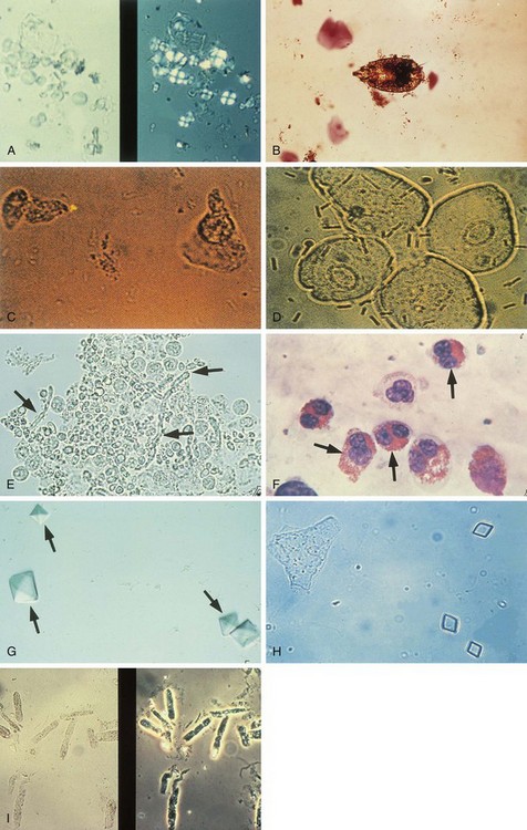
Meticulous examination of the urine sediment begins with centrifugation of 10 mL of freshly voided urine for 5 to 7 minutes at 3000 rpm. Such standardized preparation enables semiquantitative comparison of sequential samples in individual patients and often provides invaluable information concerning the etiology of numerous renal disorders as well as the anatomic location of hematuria or pyuria. Abnormalities commonly encountered on microscopy are shown in Figure 13-1. Dysmorphic RBCs, best seen in uncentrifuged urine by phase microscopy, are usually of glomerular origin as opposed to urologic origin. White blood cells (WBCs) coated with antibody tend to become agglutinated or clumped. Such clumped WBCs are indicative of pyelonephritis or interstitial nephritis rather than cystitis. The presence of several tubular epithelial cells is abnormal.


image

Figure 13-1 A, Left, Starch granules commonly seen in urine. Notice their variable size (7 to 40 µm), irregular shape with rounded corners, and central indentation. Right, “Maltese cross” appearance can be seen with polarized light. Talc particles have a similar appearance. Do not confuse these with lipid droplets seen in children with nephrotic syndrome and lipiduria, which are spherical with clear centers and usually measure less than 7 µm across. B, Mites (shown), pinworm ova (Enterobius vermicularis), or vegetable fibers and other fecal artifacts may be washed into the urine from the urethra or vaginal introitus. C, Tubular epithelial cells in a child with toxic tubulopathy due to amphotericin toxicity. D, Transitional epithelial cells arising from the renal pelvis, ureter, or bladder may appear “pear shaped” and can be confused with tubular epithelial cells as seen in C. Transitional cells are much larger in size, with smaller distinctive nuclei. These cell types may be found in very small numbers in healthy children. E, Yeast hyphae (arrows) and leukocytes in an immunosuppressed child. F, Wright-Giemsa staining showing eosinophiluria (arrows) associated with tubulointerstitial nephritis and acute renal failure in a child with hypersensitivity to methicillin. G, Calcium oxalate crystals (arrows), usually seen in acid urine, with characteristic octahedral or star-shaped pattern in a child with hematuria associated with hypercalciuria. H, Four-sided form of uric acid crystals; other forms include amorphous, six-sided bipyramidal or whetstone crystals. I, Coarse granular casts composed of degenerated cellular elements in a child with chronic IgA nephritis. Notice the typical parallel borders and refractivity of casts in general.


Scanning of the entire slide may be needed to detect small numbers of casts. Although this is time-consuming, it is a very rewarding practice because the presence of casts is indicative of intrinsic renal injury and may spare the child unnecessary imaging studies. Note that formation of tubular casts is aided by diminished urine flow, high urinary solute concentration, and the hyaline matrix of plasma- and tubule-derived protein in which cells become embedded.


Hyaline casts form on polymerization of Tamm-Horsfall glycoprotein, which is a well-characterized molecule uniquely found in the ascending limb of Henle and distal convoluted tubule. Small quantities of hyaline casts may be found in healthy individuals, but larger numbers of hyaline casts or those containing WBCs or RBCs are considered pathologic. Muddy-brown granular or epithelial cell casts are seen in acute tubular necrosis. Granular casts form from disintegration of cellular elements. These are always pathologic and may be found in most cases of glomerulonephritis. Wide casts may derive from dilated tubules, which occur in individuals with advanced renal disease.


Crystals are best identified in fresh urine that is of near-body temperature. Uric acid crystals and urates are found in urine with a pH below 6.5, whereas phosphates precipitate in urine with a pH above 6.5. Both urates and phosphates precipitate when the urine specimen is refrigerated. Crystals may be found in healthy individuals and in patients with urolithiasis or hyperuricemia, or in individuals with a specific drug intake or poisoning (e.g., ethylene glycol). Exceptions include flat hexagonal crystals, which are pathognomonic of cystinuria.


Hematuria


Isolated gross or microscopic hematuria is probably the most common symptom prompting nephrologic assessment in children. Many such children have asymptomatic microscopic hematuria often detected during routine office visits or physical examinations required before participation in sport activities. Because of the large number of conditions associated with persistent hematuria in children, several algorithms have been devised to aid in the systematic evaluation of this condition (Fig. 13-2).
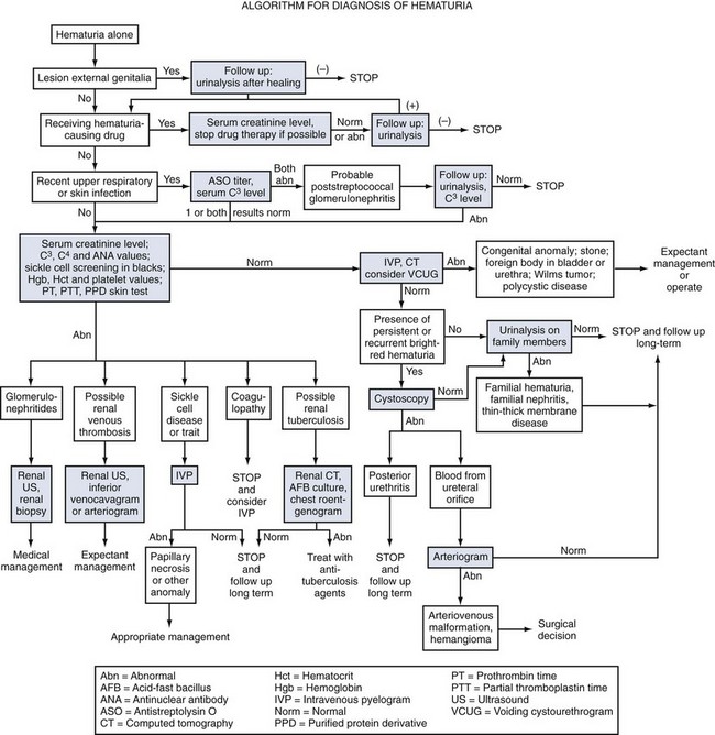


History and clinical symptoms may point toward trauma, viral or bacterial cystitis, drug-induced hematuria, or other causes. Detection of the most common causes of hematuria, including glomerulonephritis or UTI, can be readily achieved by the finding of cellular casts in a carefully performed examination of the urinary sediment or by appropriate bacterial cultures. Moreover, the absence of red blood cells in a child with positive o-toluidine reagent color change on dipstick testing may lead to the correct diagnosis of conditions associated with rhabdomyolysis or hemolysis. Once these simple measures are undertaken, biochemical techniques are used to investigate renal function and hyperexcretion of metabolites resulting in nephrolithiasis, hemoglobinopathies, or bleeding diathesis, or to perform immunologic assessment of an underlying glomerulonephritis. Measurement of calcium and creatinine concentrations in a single voided urine sample should also be included in the minimal initial assessment of asymptomatic hematuria, because hypercalciuria is found in a large proportion of such children. Identification of possible disorders by such methods may help determine the need for further assessment. Thus, the finding of a nephritic sediment obviates the need for any radiologic procedures, whereas the presence of recurrent well-documented UTIs may be an indication for imaging procedures. In the absence of any physical signs, such as an abdominal mass to suggest Wilms tumor or neuroblastoma, malignancies of the kidney or urinary tract rarely present with isolated gross or microscopic hematuria. Renal ultrasonography coupled with Doppler evaluation of the renal vessels is useful in screening for the presence of tumor, polycystic kidney disease, or renal venous thrombosis in infants. Computed tomography or nuclear magnetic resonance techniques may provide detailed anatomic resolution of such masses.


Invasive cystography or arteriography is rarely necessary in the evaluation of structural lesions underlying isolated hematuria in children. A technetium-99m (99mTc)-dimercaptosuccinic acid renal scan may disclose renal scars suggestive of chronic pyelonephritis in children with or without vesicoureteral reflux. Last, a renal biopsy may be helpful in making a definitive diagnosis in cases of suspected renal parenchymal disease manifested by hematuria.


Glomerular Disorders


Nephritis and Nephrosis


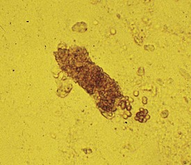
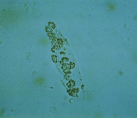
In children suspected of having a glomerular disease, the urinary sediment can provide important clues that may expedite the diagnosis and help formulate therapeutic plans. A classic example of nephritic syndrome is that of acute postinfectious glomerulonephritis, in which the urinalysis reveals variable levels of proteinuria; and granular, red (Fig. 13-3) and, less frequently, white (Fig. 13-4) blood cell casts. Acute poststreptococcal glomerulonephritis is the most common and classic example of postinfectious glomerulonephritis, although other preceding infections can cause glomerulonephritis. On the other hand, the urine of children with classic nephrotic syndrome, such as minimal change disease, shows heavy proteinuria (>40 mg/m2/hour or 1000 mg/m2/day), free fat droplets and oval fat bodies, and little or no hematuria or other sediment abnormalities.


Acute Glomerulonephritis


Acute glomerulonephritis is characterized by the sudden onset of painless dark, cola- or tea-colored urine; proteinuria; and cellular casts on urine microscopy. Edema, hypertension, and renal insufficiency are common clinical findings and together constitute the nephritic syndrome, which implies the presence of renal inflammation. The most common cause of acute glomerulonephritis in children is acute poststreptococcal glomerulonephritis. This condition follows pharyngitis, otitis media, or pyoderma caused by one of about 10 nephritogenic strains of group A β-hemolytic streptococci. Only about 15% of children infected with these strains manifest clinical symptoms to suggest nephritis. The disorder most frequently affects children ages 4 to 12 years and is more common in males. Both humoral (immune complexes with predilection for glomeruli) and cellular factors have been implicated in the pathogenesis of this disorder. Most symptoms and signs resolve within a few weeks, although microscopic hematuria may last for up to 1 year. Complete recovery of renal function is the rule, with only a few children progressing to renal failure. Other conditions that may present as acute glomerulonephritis include IgA nephritis and Henoch-Schönlein purpura.


In some instances, these disorders may have an aggressive clinical course characterized by oliguria, hypertension, and rapid reduction in glomerular filtration rate, in which case the designation of rapidly progressive glomerulonephritis (RPGN) is given. Renal biopsy in such patients often demonstrates cellular or acellular crescents and inflammatory infiltrates.


In addition to the clinical symptoms, anti-nuclear antibody titer, anti–streptolysin O titer, anti-neutrophilic cytoplasmic antibody titer, serum immunoglobulin concentrations, and C3 and C4 levels are often helpful in differentiating several of the glomerulonephritides. Complement levels are particularly helpful because only a few of these conditions are associated with depressed levels. The most common of these conditions in pediatrics include acute poststreptococcal glomerulonephritis, lupus nephritis, membranoproliferative glomerulonephritis, and glomerulonephritis associated with chronic indolent infections (ventriculoperitoneal shunt infection, endocarditis, and occult abscess). In poststreptococcal glomerulonephritis, the C3 levels are only transiently reduced and return to normal concentrations within 8 weeks after onset of the renal symptoms.


A typical presentation is that of a child 3 to 10 years of age, whose symptoms during the preceding few days have included mild periorbital edema, headache, and decreasing urine output. The urine is described as being smoky or tea colored. Medical history reveals that 2 weeks earlier the child experienced a febrile illness with painful pharyngitis for which he received no medical attention. Clinical examination reveals a blood pressure of 140/105 mm Hg, mild periorbital edema, and tenderness on palpation of the kidneys. A urinalysis shows the following values: 2+ protein, 3+ blood, and an SG of 1.020. Red blood cell casts (see Fig. 13-3) are seen on urinalysis. Laboratory studies are consistent with mild renal insufficiency and also revealed a protein excretion rate of 1.1 g/24 hours, a low plasma C3 level, and elevated streptozyme and anti-DNase B titers, evidence that strongly implicates a streptococcal infection in the pathogenesis of the glomerulonephritis.


Chronic Glomerulonephritis


White blood cell casts may be seen in the urine sediment of patients with acute or chronic glomerulonephritis, vasculitis, pyelonephritis, and other disorders resulting in tubulointerstitial nephritis. The cast shown in Figure 13-4 occurred in a child with systemic lupus erythematosus whose only symptom was mild back pain. Urinalysis demonstrated 2+ protein, microhematuria, pyuria without bacteria, and red and white blood cell casts. Diagnosis was confirmed by immunologic findings including low serum C3 and C4 levels, high anti-nuclear antibody titer, and antibodies against double-stranded DNA. Renal biopsy revealed diffuse proliferative lupus nephritis. Several acute glomerular syndromes may progress to chronic glomerulonephritis. In the final stages, many such patients develop hypertension and severe renal failure (uremia). On renal ultrasonography, the kidneys appear small and hyperechoic due to fibrosis.


Henoch-Schönlein Purpura


Three weeks after a respiratory infection, a 2-year-old boy experienced symptoms of generalized malaise, abdominal pain, periorbital edema, and difficulty walking “as if his legs were hurting.” One day later he developed an ecchymotic, purpuric rash, the characteristic clinical manifestation of Henoch-Schönlein purpura (HSP). The rash covered the extensor surfaces of the extremities and the buttocks but spared the trunk. Individual lesions faded over 1 week, but new lesions appeared or recurred over several weeks. Other cutaneous manifestations of the vasculitic lesions in this disorder are shown in Figures 13-5 and 13-6.
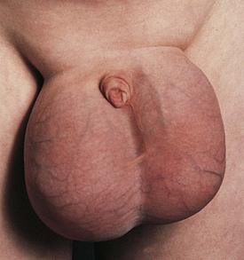


Some patients initially develop an urticarial-type eruption that subsequently becomes macular or maculopapular. On occasion, younger patients develop an angioneurotic-like edema of the scalp, face, or dorsum of the hands or feet. Of children with HSP, 90% have a prodrome consisting of an upper respiratory infection 1 to 3 weeks before the onset of symptoms, and 80% have melena, hematemesis, and/or arthritis mostly involving the ankles and knees. About half of the patients have renal involvement ranging from simple microscopic hematuria and a variable degree of proteinuria, to oliguria and renal failure. In contrast to adults, use of multiple medications is rarely related to the onset of this condition in children.


There are no distinct biochemical features of this disorder. Leukocytosis and an elevated serum IgA level may be present. In the absence of severe proteinuria, hypoalbuminemia and edema are often a result of protein-losing enteropathy. Platelet counts and coagulation studies are normal. The skin rash is essential for the diagnosis of HSP because the renal abnormalities may otherwise closely resemble a similar disorder known as IgA nephropathy (Berger disease).


Nephrotic Syndrome


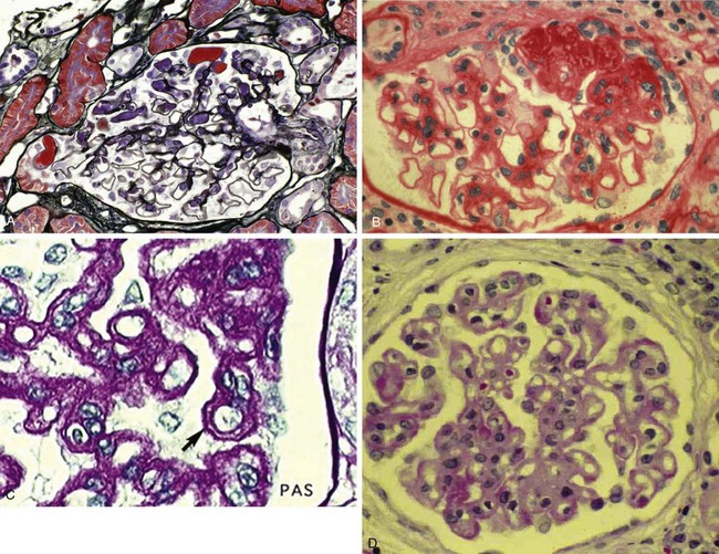
Nephrotic syndrome (NS) is defined as any renal disorder resulting in marked proteinuria (≥40 mg/m2/hour, ≥1000 mg/m2/day, or spot urine protein-to-creatinine ratio > 2.0), leading to hypoalbuminemia, hypercholesterolemia, and edema. Generalized edema and rapid weight gain are characteristic features of this condition, with the former showing a predilection for the eyelids, pleural spaces, abdomen, scrotum, and lower extremities (Figs. 13-7 and 13-8). Although edema usually provokes few complaints, at times it may be disfiguring, and it may produce skin induration and breakdown, or interference with respiratory, genitourinary, or gastrointestinal function. Children with NS rarely have an underlying systemic illness or a history of drug intake and thus are designated as having primary or idiopathic NS. The most common histopathologic entities of noninflammatory glomerular disorders associated with primary NS of childhood include minimal change disease (MCD), focal segmental glomerulosclerosis (FSGS), and membranoproliferative glomerulonephritis (MPGN); membranous glomerulopathy (MGP) is also encountered in older children and in adults. Examples of the pathology of these disorders are depicted in Figure 13-9, A-D.


Lower levels of proteinuria, termed nephrotic-range proteinuria, may also develop in children with acute poststreptococcal glomerulonephritis, HSP, IgA nephritis, or systemic lupus erythematosus, as well as rare patients treated with nonsteroidal antiinflammatory agents and other drugs. Such individuals do not develop the other features of NS.


The clinicopathologic correlation between NS associated with MCD and its favorable response to steroids has become increasingly less important over the past three decades. For example, a landmark multicenter study of childhood NS from 1967 to 1976 (International Study of Kidney Disease in Children, 1978, 1981) found that nearly 80% of all children with NS had MCD (e.g., normal pathology under light microscopy and podocyte foot process fusion seen on electron microscopy), and the chances of undergoing remission by the use of corticosteroids was 93% to 98%. In contrast, FSGS accounted for 5% to 7% of the cases of NS and only 17% to 30% underwent remission after 1 month of treatment with an adequate daily dose of steroids. Also, the same study determined that whereas multiple relapses are common in children with MCD, late resistance to steroids occurred in only 3.3%. More recent experience has challenged this paradigm. The proportion of all children presenting with NS who respond to steroids is perhaps less than 55% regardless of the underlying histopathology, and late resistance to steroids in those with MCD may exceed 20% of cases. Consequently, over the past 15 to 20 years clinicians use the terms “steroid sensitive” (SSNS), “steroid dependent” (SDNS), and “steroid resistant” (SRNS) to classify individuals with NS, rather than rely solely on the renal biopsy diagnosis to guide management decisions and predict prognosis. It should be noted that in addition to children with SRNS, those with SSNS who develop relapses while still taking steroids, and those with SDNS, have a high likelihood of developing SRNS. In children with SDNS and especially in those with SRNS, treatment often evolves by trial and error.


With the exception of some relatively rare genetic disorders leading to NS, the etiology of idiopathic or primary NS is unknown. Several genetic and acquired derangements in podocytes and their epithelial foot processes lining the glomerular capillaries have been identified, and are collectively known as podocytopathies. However, the triggering stimulus of primary NS is unknown. Activation of various immunologic mechanisms, possibly stimulated by infectious agents, is implicated. Several cytokine derangements and vascular permeability factors may also play a role in the development of proteinuria. Regardless of the underlying etiology, both the duration and the magnitude of proteinuria correlate well with the development of renal injury. It is hypothesized that the protein overload overwhelms the capacity of proximal tubular epithelial cells to reabsorb and process proteins escaping the glomerular barrier, resulting in accelerated apoptosis or direct injury to these cells, ultimately leading to interstitial fibrosis. Thus, achieving remission or reducing the amount of proteinuria is a major goal of medical management.


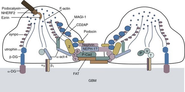
Although genetic mutations of laminin B2 and B3 chain genes and other structural proteins of the glomerular basement membrane can lead to NS, genetic mutations leading to derangements in transcription of several proteins that maintain the structure and function of the slit diaphragm of podocytes are much more common and underscore the importance of this structure as the main barrier to proteinuria (Fig. 13-10). The Finnish-type congenital NS is the prototype of a genetic mutation of nephrin and accounts for 6.25% of children with SRNS. Podocin mutations account for most familial cases of FSGS and of steroid-resistant cases (18%), especially among African-American children. Actinin IV mutations are autosomal dominant and cause a cytoplasmic defect in the podocytes (not in the slit pore diaphragm). African Americans with a form of the specific MYH9 polymorphism that encodes myosin heavy chain IIA have a much higher risk for developing FSGS in association with human immunodeficiency virus (HIV) infection even during the first few months of life. This may lead to poor association of myosin and actin and disruption of the podocyte cytoskeleton.


However, although genetic mutations point to the importance of aberrant protein transcription in the development of proteinuria and NS, the rising incidence of SRNS and FSGS, especially among African-American children in the United States, suggests an important contribution of environmental rather than genetic influences in triggering primary NS. Thus, acquired and therefore potentially reversible disorders of podocyte function account for the majority of cases of NS. One hypothesis implicates an alteration in the cathepsin L proteolytic pathway of podocytes so as to change cell metabolism and result in proteinuria (Kistler et al, 2010). In fact, a major benefit of cyclosporine use in primary NS may be mediated by direct protection of the podocyte actin cytoskeleton from cathepsin-mediated injury rather than from an immunosuppressive action. Such an understanding may lead to podocyte-targeted therapies. Similarly, deletion of CD2-associated protein (CD2AP), a protein that interacts with nephrin and is essential in maintaining slit diaphragm integrity, leads to NS. Genetic traits may also be important in conferring responsiveness to steroids and calcineurin inhibitors by reversing aberrant mechanisms in podocytes.


Management of SDNS and of SRNS is one of the major challenges in pediatric nephrology today. Management is limited by the lack of clear understanding of the pathophysiology of the underlying disorder and by a dearth of large-scale, placebo-controlled studies. All therapies have potential serious adverse effects. Transplantation has been a successful option in kidney failure; however, in many patients, NS may recur.


Cyclophosphamide has been recommended as the first-line drug for SDNS and for SRNS and is used for a 3-month period. Because a large number of children with biopsy-confirmed FSGS are unresponsive to cyclophosphamide, calcineurin inhibitors such as tacrolimus or cyclosporine are increasingly becoming the first-line drug for FSGS, thereby sparing unnecessary gonadal and other toxicity. Mycophenolate mofetil (MMF) is often added to help maintain remission while allowing reduction of the dosages of steroids and limiting the nephrotoxicity associated with long-term administration of calcineurin inhibitors. Angiotensin receptor blockers and angiotensin-converting enzyme inhibitors are often employed as adjunct antiproteinuric and/or antihypertensive agents in children with SRNS. Plasmapheresis to remove a putative vascular permeability factor has been applied in some individuals with SRNS associated with FSGS, with variable success.


In children with SRNS refractory to the previously described agents, rituximab, an anti-CD20 antibody directed against B cells that may have a regulatory effect on T cells, may be effective. Theoretically, bortezomib, a proteasome inhibitor that may limit antibody synthesis by plasma cells, may also have a benefit in SRNS. However, the efficacy and safety of these agents have not been adequately investigated in the setting of NS.


Steroids are also useful in managing less common causes of NS such as MPGN (see Fig. 13-9, C), or steroids alternating with cyclophosphamide for 6 months in adolescents with membranous glomerulopathy (see Fig. 13-9, D).


Because agents used to manage NS may have significant short- and long-term toxicity, investigations are currently focusing on the identification of blood and urine biomarkers as “surrogate markers” of the underlying histopathology and steroid resistance, thus permitting the earlier use of other, more effective agents, as well as monitoring of response to treatment. For example, the urine level of CD80 differentiation antigen present in podocytes and in antigen-presenting cells rises in MCD during relapses of NS in children but falls while they are in remission. CD80 is not present in the urine of children with FSGS or in healthy children and may be up-regulated by T cell–derived cytokine interleukin (IL)-13. Serum IgE also increases in children during relapses of MCD. Another study showed a rise in CD19+ lymphocyte count in association with relapses of SDNS or calcineurin-sensitive NS, suggesting an important role of this cell subset in NS.


Pediatric Nephrolithiasis


The diagnosis of nephrolithiasis should be entertained in any child with acute onset of costovertebral angle or abdominal/flank colicky pain. In younger children, renal colic is poorly localized and is often described as diffuse abdominal pain. Small stones may produce no pain and may be detected only after an episode of painless gross hematuria, pyuria, or UTI. Thus, a strong index of suspicion is required on the part of the clinician so that appropriate diagnostic studies are undertaken. Although dietary phytate is a more common cause of endemic stones in the Far East and UTI is more common in Europe, metabolic disorders predominate in children with nephrolithiasis in the United States. Relatively few children pass gravel or stones, and the kind of crystals found in the urine are rarely of diagnostic value. The clinical history and laboratory evaluation often reveal the cause of the stones. Direct chemical analysis of the calculus may also disclose the composition of the stone. One diagnostic approach to pediatric nephrolithiasis is shown in Table 13-2.


Table 13-2 Evaluation of Nephrolithiasis


Clinical History

< div class='tao-gold-member'>

Stay updated, free articles. Join our Telegram channel

Jul 11, 2016 | Posted by in PEDIATRICS | Comments Off on Nephrology

Full access? Get Clinical Tree

Get Clinical Tree app for offline access