Chapter 11 Miscarriage and abortion
Data from several countries estimate that between 10 and 20% of clinically diagnosed pregnancies end in miscarriage. Miscarriage is more frequent among women over the age of 30 and increases further among women over the age of 35; the risk being nine times greater than for women aged 20–29. Paternal age over 40 also increases the risk, albeit not as strongly as maternal age. The risk also increases in frequency with increasing gravidity: 6% of first or second pregnancies terminate as a miscarriage; with third and subsequent pregnancies the rate increases to 16%.
AETIOLOGY OF SPONTANEOUS MISCARRIAGE
The causes of miscarriage are:
Implantation
Implantation occurs 8–10 days after ovulation in most healthy pregnancies. The proportion ending in early loss increases when implantation is later. A refractory period after the time of uterine receptivity may provide a natural mechanism that eliminates impaired embryos. In 20% of miscarriages the trophoblast has failed to implant adequately.
In the early weeks of pregnancy (0–10 weeks) ovofetal factors account for most miscarriages; in the later weeks (11–22 weeks) maternal factors become more common (Table 11.1).
Table 11.1 Aetiological factors in 5000 abortions
FACTOR | PERCENTAGE |
---|---|
Fetal or ovular | |
Defective ovofetus | 60 |
Defective implantation or activity of trophoblast | 15 |
Maternal | |
General disease | 2 |
Uterine abnormalities | 8 |
Psychosomatic | ?15 |
Ovofetal factors
Ultrasound examination of the fetus and subsequent histological examination show that in 70% of cases the fertilized ovum has failed to develop properly or the fetus is malformed. In 40% of these cases chromosomal abnormalities are the underlying cause of the miscarriage.
Maternal factors
Systemic maternal disease (e.g. systemic lupus erythematosus), and particularly maternal infections, account for 2% of miscarriages. A further 8% are associated with uterine abnormalities, such as congenital defects, uterine myomata, particularly submucous tumours, or cervical incompetence (see p. 104). Psychosomatic causes have been suggested as leading to miscarriage, but the evidence is difficult to evaluate. Women who smoke 10 cigarettes or more per day double their risk.
MECHANISMS OF MISCARRIAGE
The immediate cause of miscarriage is the partial or complete detachment of the embryo by minute haemorrhages in the decidua. As placental function fails uterine contractions begin, and the process of miscarriage is initiated. If this occurs before the eighth week the defective embryo, covered with villi and some decidua, tends to be expelled en masse (the so-called blighted ovum), although some of the products of conception may be retained either in the cavity of the uterus or in the cervix. Uterine bleeding occurs during the expulsion process.
Between the eighth and 14th weeks the above mechanism may occur or the membranes may rupture, expelling the defective fetus but failing to expel the placenta, which may protrude through the external cervical os or remain attached to the uterine wall. This type of miscarriage may be attended by considerable haemorrhage.
Between the 14th and 22nd weeks the fetus is usually expelled followed, after an interval, by the placenta. Less commonly the placenta is retained. Usually bleeding is not severe, but pain may be considerable and resemble a miniature labour.
It is clear from this description that miscarriage is attended by uterine bleeding and pain, both of varying intensity. Although miscarriage is the cause of bleeding per vaginam in early pregnancy in over 95% of cases, less common causes, such as ectopic gestation, cervical bleeding from the everted cervical epithelium or from an endocervical polyp, hydatidiform mole, and, rarely, cervical carcinoma, must be excluded.
VARIETIES OF SPONTANEOUS MISCARRIAGE
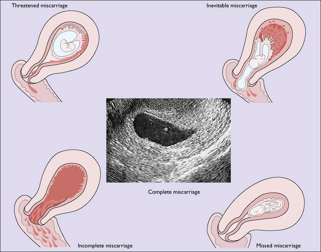
For descriptive purposes the miscarriage is classified according to the findings when the woman is first examined, but one kind may change into another if the aborting process continues. If infection complicates the miscarriage, the term septic miscarriage is used. The various types of miscarriage are shown in Figure 11.1 and each will be considered separately later.

Fig. 11.1 The types of miscarriage that may be seen. In complete miscarriage the sac contains a small amount of debris.
Threatened miscarriage
Threatened miscarriage is diagnosed when a pregnant woman develops uterine bleeding with or without painful contractions; other causes of bleeding in early pregnancy should be excluded. A vaginal examination (or vaginal speculum examination) shows that the cervix is not dilated.
A real-time pelvic ultrasound examination will clarify the diagnosis. This may show:
Only if the first finding is obtained is the diagnosis confirmed. The ultrasound finding also provides the information that the pregnancy will continue (in 98% of cases), and the patient can be reassured. If a subchorionic haematoma is detected the pregnancy should be monitored more closely, as there is a greater risk of spontaneous miscarriage, placental abruption, premature labour, intrauterine growth restriction and fetal death.
The use of ultrasound examination has meant that the treatment of threatened miscarriage has changed in recent years. It is no longer normal practice to insist that the woman remain in bed until the bleeding has ceased. However, if she feels more comfortable there, she may do so. Drugs, hormones (e.g. progesterone) and sedatives have no effect except as a placebo, and should be avoided.
Inevitable, incomplete and complete miscarriage
Miscarriage becomes inevitable if uterine bleeding is associated with strong uterine contractions that cause dilatation of the cervix. The woman complains of severe colicky uterine pains, and a vaginal examination shows a dilated cervical os with part of the conception sac bulging through. Inevitable miscarriage may follow signs of threatened miscarriage or, more commonly, starts without warning.
Soon after the onset of symptoms of inevitable miscarriage, the miscarriage occurs either completely, when all the products of conception are expelled, or incompletely when either the pregnancy sac or the placenta remains, distending the cervical canal. In most cases the miscarriage is incomplete. Unless the doctor has been able to inspect all the material expelled from the uterus, or has had an ultrasound examination that shows an empty uterus (or one containing less than 10 mm of tissues or blood clots), the miscarriage should be considered incomplete. This is treated by curettage; an alternative is to give misoprostol 400 μg 4-hourly for three doses or 800 μg as a single dose which will achieve a 60–80% complete evacuation of the uterus.

Full access? Get Clinical Tree

