12.3 Meningitis and encephalitis
Meningitis and encephalitis are both life-threatening infections that require rapid recognition and treatment for a child to survive without sequelae. They are on the differential diagnosis for all febrile ill children, particularly those with altered consciousness.
Bacterial meningitis
The improving control of leading causes of bacterial meningitis has reduced both mortality and morbidity; both vaccination and improved quality of life have contributed to dramatic declines in the incidence of the big three causes: Haemophilus influenzae type b, Neisseria meningitidis and Streptococcus pneumoniae. Consequently, the reduced familiarity of doctors with disease must be balanced by better training in the early recognition and treatment of these deadly infections.
Knowledge of local epidemiology should direct antibiotic use. Appropriate adjustments in antibiotics rely heavily on culture and sensitivity results. Improved supportive and resuscitative measures in recent decades have reduced the range of case fatality rates from 5–25% (pneumococcal meningitis and meningococcal sepsis being the most deadly) to 2%–8%. What has made the difference, at least in developed countries, includes paying close attention to:
Epidemiology
The annual incidence of bacterial meningitis is about 10–20 cases per 100 000 children aged 0–5 years, highest in infants. In Australia, the pneumococcus bacteria is now the commonest cause, and Hib immunization means that there are only a few sporadic Hib cases. Pneumococcal and meningococcal cases are sporadic too, except for occasional outbreaks of meningococcal disease. As with other serious infections, the risk of disease and its severity are determined by factors to do with the environment, the microbe and the host.
The environment
The incidence of bacterial meningitis is higher in the Indigenous and the poor, the two all too often occurring together. Prior viral respiratory infection and crowding (including school and daycare attendance) increase risk and perhaps explain why disease peaks during late winter and spring. Exposure to smokers and close personal contact is also important.
Microbe: polysaccharide capsule
A key virulence determinant is the microbe’s polysaccharide capsule. Specific human antibody to each capsule is protective.
Human genetic factors
Human genetic factors predisposing to disease are increasingly recognized, although so far they account for only a small proportion of cases. Our understanding in this area is growing rapidly.
• Defects in the various complement pathways predispose to meningococcal disease. First recognized were mutations of terminal complement components, then properdin.
• More recently, variants in mannose-binding protein and factor H binding protein have been identified as increasing the risk of meningococcal infection.
• For Hib, genetic variation in Toll-like receptor-4 and associated pathways are linked to increased risk.
• The risk of pneumococcal disease is increased by an inherited defect in interleukin-1 receptor-associated kinase-4 (IRAK-4), leading to a failure to make cytokines following stimulation with Toll-like receptor agonists such as interleukin-1β.
• Congenital absence of the spleen may have a genetic basis and predisposes to infection with encapsulated organisms.
• Immunoglobulin deficiencies (X-linked or recessive) predispose to meningitis generally, whereas recurrent meningococcal disease may be associated with immunoglobulin (Ig) M deficiency.
Other factors that increase the risk of meningitis:
• Acquired impaired immunity (e.g. human immunodeficiency virus infection) and splenectomy
• Congenital or acquired neuroanatomical defects (e.g. base of skull fracture)
• Penetrating head injuries, neurosurgical procedures
• Cerebrospinal fluid (CSF) leak
• Congenital dural defect (dermal sinus or myelomeningocele)
Aetiology
The most common pathogens in each age group are:
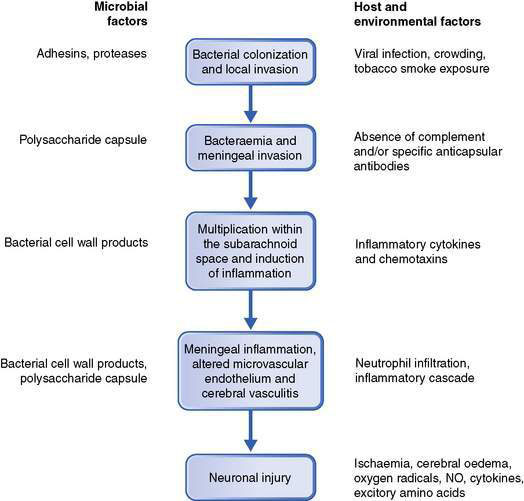
Pathogenesis
The development of bacterial meningitis usually follows: (1) colonization of the nasopharynx by encapsulated bacteria; (2) invasion of the host causing bacteraemia and then invasion of the meninges; (3) bacterial multiplication and induction of inflammation within the subarachnoid space; and (4) neuronal injury (Fig. 12.3.1).
Clinical presentations
Infants
Many symptoms and signs within this age group are non-specific:
Neck stiffness or a bulging fontanelle are more specific but may be absent, especially early in the illness.
Children aged at least 3 years
Symptoms and signs are more obvious:
• severe headache, vomiting, photophobia
• fever, neck stiffness, delirium or deteriorating consciousness.
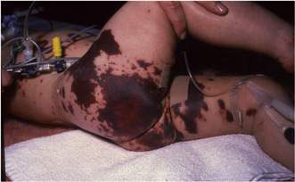
Convulsions are a presenting feature in 20–30% of infants and children with bacterial meningitis. Those with meningococcal meningitis or septicaemia may have a petechial and/or purpuric rash over the trunk, limbs and, in extremis, the face. Initially the rash may be blanching, but within hours become non-blanching purpuric lesions that may progress through dermal vascular necrosis to peripheral gangrene (Fig. 12.3.2).
Diagnosis
Lumbar puncture (LP) helps to establish and localize the infection through obtaining a sample for microscopy, culture, biochemistry and molecular diagnostic testing. Unless the child is already treated with antibiotics, LP has a very high sensitivity. Molecular diagnostic testing using polymerase chain reaction (PCR) on blood or CSF can identify the cause of meningitis in patients pretreated with antibiotics.
Owing to concern over the dangers of cerebral herniation, circulatory compromise or disseminated intravascular coagulation, in recent years LP has often been postponed; immediate collection of peripheral blood samples for diagnosis (culture, PCR) is helpful but diagnostic sensitivity may be reduced by up to 50%. The suspicion of bacterial meningitis should prompt rapid commencement of antibiotics; this is ultimately a clinical decision.
Paired serology is helpful, but only in retrospect.
• Needle aspirate from skin lesions for Gram stain and culture is useful for diagnosis in meningococcal disease.
• Suprapubic aspirate or catheter urine specimen, if less than 6 months old, may reveal dual sites of infection.
• Full blood count, including white blood cell differential, C-reactive protein or procalcitonin may all point to the seriousness of infection.
• Serum electrolytes, glucose and creatinine should be monitored and managed.
Lumbar puncture is delayed for patients with any of the following:
• absent or non-purposeful responses to pain
• focal neurological signs (or a false localizing sixth nerve palsy)
• abnormal pupil size or reaction
• decerebrate or decorticate posturing
• irregular breathing, rising blood pressure and falling pulse (despite peripheral vasoconstriction)
Such patients require intensive care management, including measures to reduce intracranial pressure. LP should still be performed, but only when the child is stable, usually within 2–3 days. Cerebral herniation occurs in about 5% of cases, with or without LP, and may account for 30% of the deaths.
The typical CSF changes in bacterial meningitis are outlined in Table 12.3.1. Organisms are often seen on Gram stain of the CSF, making a presumptive diagnosis possible.
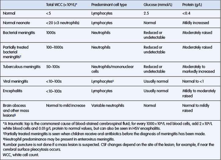
CSF examination may be difficult to interpret, especially when prior antibiotics have been given.
Parameningeal foci, such as brain abscess or subdural empyema, and tuberculous meningitis can have a similar CSF profile to partially treated bacterial meningitis, so should be in the differential diagnosis.
Cerebral imaging, by either computed tomography (CT) or magnetic resonance imaging (MRI), is not recommended routinely and is not useful for determining whether there is raised intracranial pressure. It has a role when there is concern that conditions that may mimic meningitis are present, such as intracranial mass lesions, and LP is contraindicated.
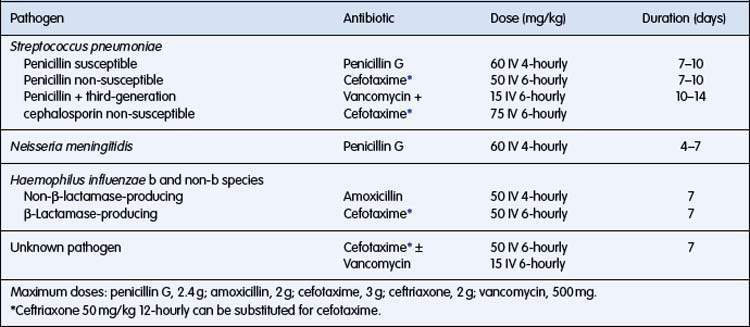
Antibiotic treatment
The antibiotics selected should cover the commonly encountered causative bacteria. As strains of S. pneumoniae resistant to penicillin and cephalosporin are relatively common in many settings, vancomycin may be used with a third-generation cephalosporin as initial empirical therapy, except in infants aged less than 3 months for whom amoxicillin should be given in addition to cover Listeria.
Subsequent adjustments depend on culture and sensitivity results.
Dosage and duration of antibiotic treatment are outlined in Table 12.3.2.
Supportive treatment
Clinical observations
Keeping a regular record of heart and respiratory rates, blood pressure, temperature and conscious state can detect important trends such as falling heart rate and rising blood pressure with rising intracranial pressure. The head circumference in infants should be measured daily.
Fluid therapy
Intravenous fluids are administered initially to restore circulating blood volume, to correct glucose or electrolyte disturbance, and to minimize the need to swallow, with its attendant risk of aspiration.
In contradiction to past recommendations, recent studies have shown that fluid restriction is not helpful for children with bacterial meningitis and, indeed, the use of maintenance fluid is associated with fewer neurological complications. Fluid administration should be adjusted according to vital signs and adequacy of circulation. The low serum sodium levels and reduced urine output that may occur in bacterial meningitis are due to fluid depletion rather than inappropriate antidiuretic hormone secretion.
Corticosteroids
Dexamethasone for treating bacterial meningitis is controversial and depends on the cause. Children with Hib meningitis have a reduced risk of deafness if dexamethasone is given prior to antibiotics. Benefit, or detriment, in children with either pneumococcal or meningococcal meningitis remains unproven; however, a recent Cochrane review showed a lower case-fatality rate and fewer long-term sequelae in a combined adult/child group of bacterial meningitis cases. A reasonable approach for bacterial meningitis of, as yet, uncertain aetiology might be to give dexamethasone 0.15 mg/kg just before the first dose of empirical antibiotics and to continue 6 hourly for 4 days, but to cease if N. meningitidis becomes the principal diagnosis.
Acute complications
During meningitis, complications from central nervous system infection or systemic effects of infection are common. Children with recurrent or protracted convulsions, circulatory instability or signs of cerebral oedema should be managed in an intensive care unit.
Convulsions
About 30% of children with bacterial meningitis have convulsions, more commonly with pneumococcal or Hib meningitis than meningococcal meningitis. Benzodiazepines such as diazepam or midazolam will control most convulsions, but if these recur or are prolonged they may raise intracranial pressure and worsen the cerebral ischaemic injury. Administration of phenytoin or phenobarbital, and mechanical ventilation may be necessary. Consider aberrations in sodium, calcium or glucose levels.
Cerebral oedema
Careful observation for signs of increased intracranial pressure (IIP) is essential. These are listed above under reasons for delaying a LP. Refer also above to section on fluid therapy. The child with suspected raised IIP should be nursed head-up and in midline, be sedated, intubated and ventilated, and given boluses of hypertonic saline or mannitol to reduce pressure. Regular oral glycerol (with or without dexamethasone) may reduce both mortality and neurological sequelae, especially in cases caused by Hib.
Circulatory shock
Approximately 5–10% of children with bacterial meningitis present in shock and initially require large-volume fluid resuscitation in 10–20-mL/kg boluses. If a second bolus is required, strong consideration should be given to elective intubation and ventilation, but in a third-world setting without ventilator support research suggests fluids be given more slowly (i.e. not by bolus).
Inotropic agents may be needed to help support the circulation.

Stay updated, free articles. Join our Telegram channel

Full access? Get Clinical Tree

