Mechanical Ventilation in Pediatric Surgical Disease
Amazingly, ventilation via tracheal cannulation was performed as early as 1543 when Vesalius demonstrated the ability to maintain the beating heart in animals with open chests.1 This technique was first applied to humans in 1780, but there was little progress in positive-pressure ventilation until the development of the Fell–O’Dwyer apparatus. This device provided translaryngeal ventilation using bellows and was first used in 1887 (Fig. 7-1).2,3 The Drinker–Shaw iron lung, which allowed piston-pump cyclic ventilation of a metal cylinder and concomitant negative-pressure ventilation, became available in 1928 and was followed by a simplified version built by Emerson in 1931.4 Such machines were the mainstays in the ventilation of victims of poliomyelitis in the 1930s through the 1950s.
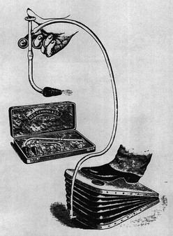
FIGURE 7-1 The Fell–O’Dwyer apparatus that was first used to perform positive-pressure ventilation in newborns. (Reprinted from Matas R. Intralaryngeal insufflation. JAMA 1900;34:1468–73.)
In the 1920s, the technique of tracheal intubation was refined by Magill and Rowbotham.5,6 In World War II, the Bennett valve, which allowed cyclic application of high pressure, was devised to allow pilots to tolerate high-altitude bombing missions.7 Concomitantly, the use of translaryngeal intubation and mechanical ventilation became common in the operating room as well as in the treatment of respiratory insufficiency. However, application of mechanical ventilation to newborns, both in the operating room and in the intensive care unit (ICU), lagged behind that for children and adults.
The use of positive-pressure mechanical ventilation in the management of respiratory distress syndrome (RDS) was described in 1962.8 It was the unfortunate death of Patrick Bouvier Kennedy at 32 weeks gestation in 1963 that resulted in additional National Institutes of Health (NIH) funding for research in the management of newborns with respiratory failure.9 The discovery of surfactant deficiency as the etiology of RDS in 1959, the ability to provide positive-pressure ventilation in newborns with respiratory insufficiency in 1965, and demonstration of the effectiveness of continuous positive airway pressure in enhancing lung volume and ventilation in patients with RDS in 1971 set the stage for the development of continuous-flow ventilators specifically designed for neonates.10–12 The development of neonatal ICUs (NICUs), hyperalimentation, and neonatal invasive and noninvasive monitoring enhanced the care of newborns with respiratory failure and increased survival in preterm newborns from 50% in the early 1970s to more than 90% today.13
Physiology of Gas Exchange during Mechanical Ventilation
The approach to mechanical ventilation is best understood if the two variables of oxygenation and carbon dioxide elimination are considered separately.14
Carbon Dioxide Elimination
The primary purpose of ventilation is to eliminate carbon dioxide, which is accomplished by delivering tidal volume (Vt) breaths at a designated rate. The product (Vt × rate) determines the minute volume ventilation ( ). Although CO2 elimination is proportional to
  , it is, in fact, directly related to the volume of gas ventilating the alveoli because part of the
  resides in the conducting airways or in nonperfused alveoli. As such, the portion of the ventilation that does not participate in CO2 exchange is termed the dead space (Vd).15 In a patient with healthy lungs, this dead space is fixed or ‘anatomic’, and consists of about one-third of the tidal volume (i.e., Vd/Vt = 0.33). In a setting of respiratory insufficiency, the proportion of dead space (Vd/Vt) may be augmented by the presence of nonperfused alveoli and a reduction in Vt. Furthermore, dead space can unwittingly be increased through the presence of extensions of the trachea such as the endotracheal tube, a pneumotachometer to measure tidal volume, an end-tidal CO2 monitor, or an extension of the ventilator tubing beyond the ‘Y.’
Tidal volume is a function of the applied ventilator pressure and the volume/pressure relationship (compliance), which describes the ability of the lung and chest wall to distend. At the functional residual capacity (FRC), the static point of end expiration, the tendency for the lung to collapse (elastic recoil) is in balance with the forces that promote chest wall expansion.15 As each breath develops, the elastic recoil of both the lung and chest wall work in concert to oppose lung inflation. Therefore, pulmonary compliance is a function of both the lung elastic recoil (lung compliance) and that of the rib cage and diaphragm (chest wall compliance).
The compliance can be determined in a dynamic or static mode. Figure 7-2 demonstrates the dynamic volume/pressure relationship for a normal patient. Note that application of 25 cmH2O of inflating pressure (ΔP) above static FRC at positive end-expiratory pressure (PEEP) of 5 cmH2O generates a Vt of 40 mL/kg. The lung, at an inflating pressure of 30 cmH2O when compared with ambient (transpulmonary) pressure, is considered to be at total lung capacity (TLC) (Table 7-1). Note that the loop observed during both inspiration and expiration is curvilinear. This is due to the resistance that is present in the airways and describes the work required to overcome air flow resistance. As a result, at any given point of active flow, the measured pressure in the airways is higher during inspiration and lower during expiration than at the same volume under zero-flow conditions. Pulmonary compliance measurements, as well as alveolar pressure measurements, can be effectively performed only when no flow is present in the airways (zero flow), which occurs at FRC and TLC. The change observed is a volume of 40 mL/kg and pressure of 25 cmH2O or 1.6 mL/kg/cmH2O. This is termed effective compliance because it is calculated only between the two arbitrary points of end inspiration and end expiration.
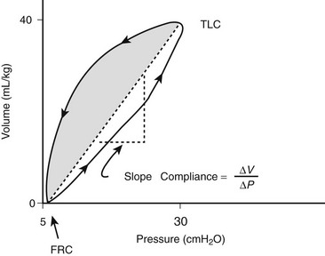
FIGURE 7-2 Dynamic pressure/volume relation and effective pulmonary compliance (Ceff) in the normal lung. The volume at 30 cmH2O is considered total lung capacity (TLC). Ceff is calculated by ΔV/ΔP. (Adapted from Bhutani VK, Sivieri EM. Physiological principles for bedside assessment of pulmonary graphics. In: Donn SM, editor. Neonatal and Pediatric Pulmonary Graphics: Principles and Applications. Armonk, NY: Futura Publishing; 1998.)
As can be seen from Figure 7-3, the volume/pressure relationship is not linear over the range of most inflating pressures when a static compliance curve is developed. Such static compliance assessments are most commonly performed via a large syringe in which aliquots of 1–2 mL/kg of oxygen, up to a total of 15–20 mL/kg, are instilled sequentially with 3 to 5-second pauses. At the end of each pause, zero-flow pressures are measured. By plotting the data, a static compliance curve can be generated. This curve demonstrates how the calculated compliance can change depending on the arbitrary points used for assessment of the effective compliance (Ceff).16
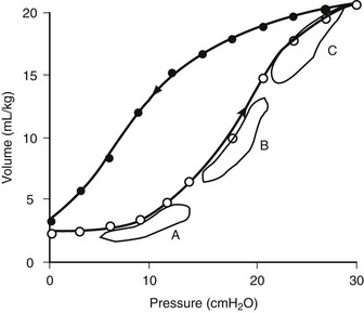
FIGURE 7-3 Static lung compliance curve in a normal lung. Effective compliance would be altered depending on whether FRC were to be at a level resulting in lung atelectasis (point A) or over distention (point C). Optimal lung mechanics are observed when FRC is set on the steepest portion of the curve (point B). (Adapted from West JB. Respiratory Physiology. Baltimore: Williams & Wilkins; 1985.)
Alternatively, the pulmonary pressure/volume relationship can be assessed by administration of a slow constant flow of gas into the lungs with simultaneous determination of airway pressure.17,18 A curve may be fitted to the data points to determine the optimal compliance and FRC.19 The compliance will change as the FRC or end-expiratory lung volume (EELV) increases or decreases. For instance, as can be seen in Figure 7-3, at low FRC (point A), atelectasis is present. A given ΔP will not optimally inflate alveoli. Likewise, at a high FRC (point C), because of air trapping or application of high PEEP, the lung is already distended. Application of the same ΔP will result only in over distention and potential lung injury with little benefit in terms of added Vt. Thus, optimal compliance is provided when the pressure/volume range is on the linear portion of the static compliance curve (point B). Clinically, the compliance at a variety of FRC or PEEP values can be monitored to establish optimal FRC.20
Finally, it is important to recognize that a portion of the Vt generated by the ventilator is actually compression of gas within both the ventilator tubing and the airways. The ratio of gas compressed in the ventilator tubing to that entering the lungs is a function of the compliance of the ventilator tubing and the lung. The compliance of the ventilator tubing is 0.3–4.5 mL/cmH2O.21 A change in pressure of 15 cmH2O in a 3 kg newborn with respiratory insufficiency and a pulmonary compliance of 0.4 mL/cmH2O/kg would result in a lung Vt of 18 mL and an impressive ventilator tubing/gas compression volume of 15 mL if the tubing compliance were 1.0 mL/cmH2O. The relative ventilator tubing/gas compression volume would not be as striking in an adult. The ventilator tubing compliance is characterized for all current ventilators and should be factored when considering Vt data. The software in many ventilators corrects for ventilator tubing compliance when displaying Vt values.
Oxygenation
In contrast to CO2 determination, oxygenation is determined by the fraction of inspired oxygen (FiO2) and the degree of lung distention or alveolar recruitment, determined by the level of PEEP and the mean airway pressure (Paw) during each ventilator cycle. If CO2 was not a competing gas at the alveolar level, oxygen within the pulmonary capillary blood would simply be replaced by that provided at the airway, as long as alveolar distention was maintained. Such apneic oxygenation has been used in conjunction with extracorporeal carbon dioxide removal (ECCO2R) or arteriovenous CO2 removal (AVCO2R), in which oxygen is delivered at the carina, whereas lung distention is maintained through application of PEEP.22,23 Under normal circumstances, however, alveolar ventilation serves to remove CO2 from the alveolus and to replenish the PO2, thereby maintaining the alveolar/pulmonary capillary blood oxygen gradient.
Rather than depending on the degree of alveolar ventilation, oxygenation predominantly is a function of the appropriate matching of pulmonary blood flow to inflated alveoli (ventilation/perfusion [ ] matching).15 In normal lungs, the PEEP should be maintained at 5 cmH2O, a pressure that allows maintenance of alveolar inflation at end expiration, balancing the lung/chest wall recoil. An FiO2 of 0.50 should be administered initially. However, one should be able to wean the FiO2 rapidly in a patient with healthy lungs and normal
  matching. Areas of ventilation but no perfusion (high
  ), such as in the setting of pulmonary embolus, do not contribute to oxygenation. Therefore, hypoxemia supervenes in this situation once the average residence time of blood in the remaining perfused pulmonary capillaries exceeds that necessary for complete oxygenation. Normal residence time is threefold that required for full oxygenation of pulmonary capillary blood.
However, the common pathophysiology observed in the setting of respiratory insufficiency is that of minimal or no ventilation, with persistent perfusion (low  ), resulting in right-to-left shunting and hypoxemia. Patients with the acute respiratory distress syndrome (ARDS) have collapse of the posterior, or dependent, regions of the lungs when supine.24,25 As the majority of blood flow is distributed to these dependent regions, one can easily imagine the limited oxygen transfer and large shunt secondary to
  mismatch and the resulting hypoxemia that occurs in patients with ARDS. Attempts to inflate the alveoli in these regions, such as with the application of increased PEEP, can reduce
  mismatch and enhance oxygenation.
where FiO2 is the fraction of inspired oxygen, PB is the barometric pressure, PH2O is the partial pressure of water, and RQ is the respiratory quotient or the ratio of CO2 production (VCO2) to oxygen consumption (VO2).
where CvO2, CaO2, and CiO2 are the oxygen contents of venous, arterial, and expected pulmonary capillary blood, respectively.
where Paw represents the mean airway pressure.15
Note that the contribution of the PaO2 to DO2 is minimal and may be disregarded in most circumstances. If the hemoglobin concentration of the blood is normal (15 g/dL) and the hemoglobin is fully saturated with oxygen, the amount of oxygen bound to hemoglobin is 20.4 mL/dL (Fig. 7-4). In addition, approximately 0.3 mL of oxygen is physically dissolved in each deciliter of plasma, which makes the oxygen content of normal arterial blood equal to approximately 20.7 mL O2/dL. Similar calculations reveal that the normal venous blood oxygen content is approximately 15 mL O2/dL.
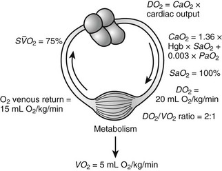
FIGURE 7-4 Oxygen consumption (VO2) and delivery (DO2) relations. (Adapted with permission from Hirschl RB. Oxygen delivery in the pediatric surgical patients. Opin Pediatr 1994;6:341–7.)
Typically, DO2 is four to five times greater than the associated oxygen consumption (VO2). As DO2 increases or VO2 decreases, more oxygen remains in the venous blood. The result is an increase in the oxygen hemoglobin saturation in the mixed venous pulmonary artery blood ( ). In contrast, if the DO2 decreases or VO2 increases, relatively more oxygen is extracted from the blood, and therefore less oxygen remains in the venous blood. A decrease in
  is the result. In general, the
  serves as an excellent monitor of oxygen kinetics because it specifically assesses the adequacy of DO2 in relation to DO2 (DO2/VO2 ratio) (Fig. 7-5).26 Many pulmonary arterial catheters contain fiber optic bundles that provide continuous mixed venous oximetry data. Such data provides a means for assessing the adequacy of DO2, rapid assessment of the response to interventions such as mechanical ventilation, and cost savings due to a diminished need for sequential blood gas monitoring.26,27 If a pulmonary artery catheter is unavailable, the central venous oxygen saturation (
 ) may serve as a surrogate of the
  .28
.28
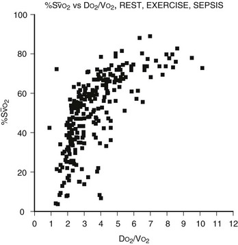
FIGURE 7-5 The relation of the mixed venous oxygen saturation (SO2) and the ratio of oxygen delivery to oxygen consumption (DO2/VO2) in normal eumetabolic, hypermetabolic septic, and hypermetabolic exercising canines. (Reprinted from Hirschl RB. Cardiopulmonary Critical Care and Shock: Surgery of Infants and Children: Scientific Principles and Practice. Philadelphia: Lippincott-Raven; 1997.)
Four factors are manipulated in an attempt to improve the DO2/VO2 ratio: cardiac output, hemoglobin concentration, SaO2, and VO2. The result of various interventions designed to increase cardiac output, such as volume administration, infusion of inotropic agents, administration of afterload-reducing drugs, and correction of acid–base abnormalities, can be assessed by the effect on the  . One of the most efficient ways to enhance DO2 is to increase the oxygen-carrying capacity of the blood. For instance, an increase in hemoglobin from 7.5 g/dL to 15 g/dL will be associated with a twofold increase in DO2 at constant cardiac output. However, blood viscosity is also increased with blood transfusion, which may result in a reduction in cardiac output.29 The SaO2 can often be enhanced through application of supplemental oxygen and mechanical ventilation.
Assessment of the ‘best PEEP’ identifies the level at which DO2 and  are optimal without compromising compliance.30,31 Evaluation of the best PEEP should be performed in any patient requiring an FiO2 greater than 0.60 and may be determined by continuous monitoring of the
  as the PEEP is sequentially increased from 5 cmH2O to 15 cmH2O over a short period. The point at which the
  is maximal indicates optimal DO2. The use of PEEP with mechanical ventilation is limited, however, by the adverse effects observed on cardiac output, the effect of barotrauma, and the risk for ventilator-induced lung injury with application of peak inspiratory pressures greater than 30–40 cmH2O.32,33 Furthermore, oxygen consumption can be elevated secondary to sepsis, burns, agitation, seizures, hyperthermia, hyperthyroidism, and increased catecholamine production or infusion. A number of interventions may be applied to reduce VO2, such as sedation and mechanical ventilation. Paralysis may enhance the effectiveness of mechanical ventilation while simultaneously reducing VO2.34,35 In the appropriate setting, hypothermia may be induced with an associated reduction of 7% in VO2 with each 1°C decrease in core temperature.36
Mechanical Ventilation
The Mechanical Ventilator and Its Components
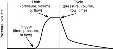
The ventilator must overcome the pressure generated by the elastic recoil of the lung at end inspiration plus the resistance to flow at the airway. To do so, most ventilators in the ICU are pneumatically powered by gas pressurized at 50 pounds per square inch (psi). Microprocessor controls allow accurate management of proportional solenoid-driven valves, which carefully control infusion of a blend of air or oxygen into the ventilator circuit while simultaneously opening and closing an expiratory valve.37 Additional components of a ventilator include a bacterial filter, a pneumotachometer, a humidifier, a heater/thermostat, an oxygen analyzer, and a pressure manometer. A chamber for nebulizing drugs is usually incorporated into the inspiratory circuit. The Vt is not usually measured directly. Rather, flow is assessed as a function of time, thereby allowing calculation of Vt. The modes of ventilation are characterized by three variables that affect patient and ventilator synchrony or interaction: the parameter used to initiate or ‘trigger’ a breath, the parameter used to ‘limit’ the size of the breath, and the parameter used to terminate inspiration or ‘cycle’ the breath (Fig. 7-6).38
The magnitude of the breath is controlled or limited by one of three variables: pressure, volume, or flow. When a breath is volume, pressure, or flow ‘controlled,’ it indicates that inspiration concludes once the limiting variable is reached. Pressure-controlled or pressure-limited modes are the most popular for all age groups, although volume-control ventilation may be of advantage in preterm newborns.39,40 In the pressure modes, the respiratory rate, the inspiratory gas flow, the PEEP level, the inspiratory/expiratory (I/E) ratio, and the Paw are determined. The ventilator infuses gas until the desired peak inspiratory pressure (PIP) is provided. Zero-flow conditions are realized at end inspiration during pressure-limited ventilation. Therefore, in this mode, PIP is frequently equivalent to end-inspiratory pressure (EIP) or plateau pressure.
In many ventilators, the gas flow rate is fixed, although some ventilators allow manipulation of the flow rate and therefore the rate of positive-pressure development. Those with rapid flow rates will provide rapid ascent of pressure to the preset maximum, where it will remain for the duration of the inspiratory phase. This ‘square wave’ pressure pattern results in decelerating flow during inspiration (Fig. 7-7). Airway pressure is ‘front loaded,’ which increases Paw, alveolar volume, and oxygenation without increasing PIP.41 However, one of the biggest advantages of pressure-controlled or pressure-limited ventilation is the ability to avoid lung over-distention and barotrauma/volutrauma (discussed later). The disadvantage of pressure-controlled or pressure-limited ventilation is that delivered volume varies with airway resistance and pulmonary compliance, and may be reduced when short inspiratory times are applied (Fig. 7-8).42 For this reason, both Vt and  must be monitored carefully.
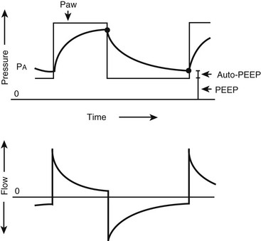
FIGURE 7-7 Pressure and flow waveforms during pressure-limited, time-cycled ventilation. Decelerating flow is applied, which ‘front loads’ the pressure during inspiration. Auto-positive end-expiratory pressure is present when the expiratory time is inadequate for complete expiration. (Reprinted from Marini JJ. New options for the ventilatory management of acute lung injury. New Horiz 1993;1:489–503.)
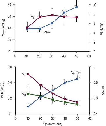
FIGURE 7-8 Effect of rate on tidal volume (Vd/Vt) and minute ventilation (VE) during pressure-limited ventilation. Note that VE remains unchanged above 20 breaths/min. Simultaneously, Vd/Vt and PaCO2 increase, despite an increase in respiratory rate. (Reprinted from Nahum A, Burke WC, Ravenscraft SA. Lung mechanics and gas exchange during pressure-control ventilation in dogs: Augmentation of CO2 elimination by an intratracheal catheter. Am Rev Respir Dis 1992;146:965–73.)
Volume-controlled or volume-limited ventilation requires delineation of the Vt, respiratory rate, and inspiratory gas flow. Gas will be inspired until the preset Vt is attained. The volume will remain constant despite changes in pulmonary mechanics, although the resulting EIP and PIP may be altered. Flow-controlled or flow-limited ventilation is similar in many respects to volume-controlled or volume-limited ventilation. A flow pattern is predetermined, which effectively results in a fixed volume as the limiting component of inspiration.
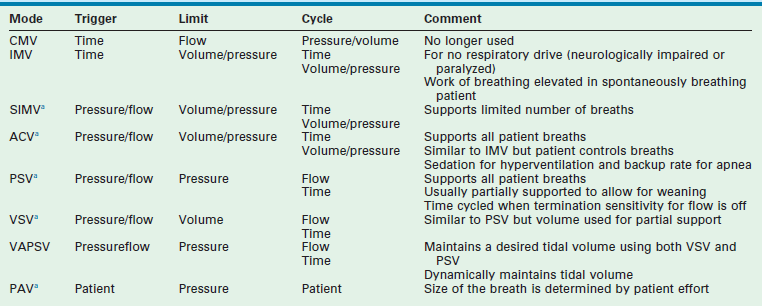
Modes of Ventilation (Table 7-2)
Intermittent Mandatory Ventilation
Intermittent mandatory ventilation (IMV) is time triggered, volume or pressure limited, and either time, volume, or pressure cycled. A rate is set, as is a volume or pressure parameter. Additional inspired gas is provided by the ventilator to support spontaneous breathing when additional breaths are desired. The difference between CMV and IMV is that, in the IMV mode, inspired gases are provided to the patient during spontaneous breaths.43 IMV is useful in patients who do not have respiratory drive such as those who are neurologically impaired or pharmacologically paralyzed. Work of breathing is still elevated with this mode in the awake and spontaneously breathing patient.
Synchronized Intermittent Mandatory Ventilation
In the SIMV mode, the ventilator synchronizes IMV breaths with the patient’s spontaneous breaths (Fig. 7-9). Small, patient-initiated negative deflections in airway pressure (pressure triggered) or decreases in the constant ventilator gas flow (bias flow) passing through the exhalation valve (flow triggered) provide a signal to the ventilator that a patient breath has been initiated. Ventilated breaths are timed with the patient’s spontaneous respiration, but the number of supported breaths each minute is predetermined and remains constant. Additional constant inspired gas flow is provided for use during any other spontaneous breaths. Advances in neonatal ventilators have provided the means for detecting small alterations in bias flow. As such, flow-triggered SIMV can be applied to newborns, which appears to enhance ventilatory patterns and allows ventilation with reduced airway pressures and FiO2.44,45 SIMV may be associated with a reduction in the duration of ventilation and the incidence of air leak in newborns in general, as well as in those premature infants with bronchopulmonary dysplasia (BPD) and intraventricular hemorrhage.46,47
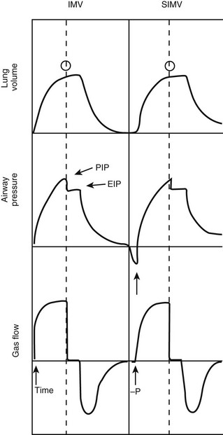
FIGURE 7-9 Pressure, volume, and flow waveforms observed during intermittent mandatory ventilation (IMV) and synchronized IMV (SIMV). In this case, an end-inspiratory pause has been added. Note the difference between peak (PIP) and end-inspiratory (EIP) or plateau pressure. Arrows, triggering variables; open circles, cycling variables. (Adapted from Bartlett RH. Use of the Mechanical Ventilator: Surgery. New York: Scientific American; 1988.)
Assist-Control Ventilation
In the spontaneously breathing patient, brain stem reflexes dependent on cerebrospinal fluid levels of CO2 and pH can be harnessed to determine the appropriate breathing rate.15 As in SIMV, with assist-control ventilation (ACV) the assisted breaths can be either pressure triggered or flow triggered. The triggering-mechanism sensitivity can be set in most ventilators. In contrast to SIMV, the ventilator supports all patient-initiated breaths. This mode is similar to IMV but allows the patient inherently to control the ventilation and minimizes patient work of breathing in adults and neonates.48,49 Occasionally, patients may hyperventilate, such as when they are agitated or have neurologic injury. Heavy sedation may be required if agitation is present. A minimal ventilator rate below the patient’s assist rate should be established in case of apnea.
Pressure Support Ventilation
Pressure support ventilation (PSV) is a pressure- or flow-triggered, pressure-limited, and flow-cycled mode of ventilation. It is similar in concept to ACV, in that mechanical support is provided for each spontaneous breath and the patient determines the ventilator rate. During each breath, inspiratory flow is applied until a predetermined pressure is attained.50 As the end of inspiration approaches, flow decreases to a level below a specified value (2–6 L/min) or a percentage of peak inspiratory flow (at 25%). At this point, inspiration terminates. Although it may apply full support, PSV is frequently used to support the patient partially by assigning a pressure limit for each breath that is less than that required for full support.51 For example, in the spontaneously breathing patient, PSV can be sequentially decreased from full support to a PSV 5–10 cmH2O above PEEP, allowing weaning while providing partial support with each breath.52,53 Thus, Vt during PSV may be dependent on patient effort. PSV provides two advantages during ventilation of spontaneously breathing patients: (1) it provides excellent support and decreases the work of breathing associated with ventilation; and (2) it lowers PIP and Paw while higher Vt and cardiac output levels may be observed.50,54,55
Volume Support Ventilation
Volume support ventilation (VSV) is similar to PSV except that a volume, rather than a pressure, is assigned to provide partial support. Automation with VSV is enhanced because there is less need for manual changes to maintain stable tidal and minute volume during weaning.56 Both VSV and PSV are equally effective at weaning infants and children from the ventilator.57
Volume-Assured Pressure Support Ventilation
Volume-assured pressure support ventilation (VAPSV) attempts to combine volume- and pressure-controlled ventilation to ensure a desired Vt within the constraints of the pressure limit. It has the advantage of maintaining inflation to a point below an injurious PIP level while maintaining Vt constant in the face of changing pulmonary mechanics. Work of breathing may be markedly decreased while Ceff is increased during VAPSV.58
Proportional Assist Ventilation
Proportional assist ventilation (PAV) is an intriguing approach in the spontaneously breathing patient. It relies on the concept that the combined pressure generated by the ventilator (Paw) and respiratory muscles (Pmus) is equivalent to that required to overcome the resistance to flow of the endotracheal tube/airways (Pres) and the tendency for the inflated lungs to collapse.59
With PAV, airway pressure generation by the ventilator is proportional at any instant to the respiratory effort (Pmus) generated by the patient. Small efforts, therefore, result in small breaths, whereas greater patient effort results in development of a greater Vt. Inspiration is patient triggered and terminates with discontinuation of patient effort. Rate, Vt, and inspiratory time are entirely patient controlled. The predominant variable controlled by the ventilator is the proportional response between Pmus and the applied ventilator pressure. This proportional assist (Paw/Pmus) can be increased until nearly all patient effort is provided by the ventilator.60 Patient work of breathing, dyspnea, and PIP are reduced.61,62 Elastance and resistance are set, as is applied PEEP. Vt is variable, and the risk of atelectasis is present. PAV produces similar gas exchange with lower airway pressures when compared with conventional ventilation in infants.63 Compared with preterm newborns being ventilated with the assist-control mode and with IMV, preterm newborns managed with PAV maintained gas exchange with lower airway pressures and a decrease in the oxygenation index by 28%.64 Chest wall dynamics also are enhanced.65 PAV represents an exciting first step in servoregulating ventilators to patient requirements. Additional studies using neutrally adjusted ventilation are also underway and certain populations (obstructive lung disease and small children) may benefit from the increased patient-ventilator synchrony.66,67
Continuous Positive Airway Pressure
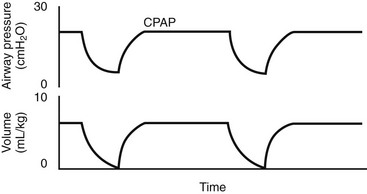
During continuous positive airway pressure (CPAP), pressures greater than those of ambient pressure are continuously applied to the airways to enhance alveolar distention and oxygenation.68 Both airway resistance and work of breathing may be substantially reduced. Since ventilation is unsupported, this mode requires that the patient provides all of the work of breathing and CPAP should be avoided in patients with hypovolemia, untreated pneumothorax, lung hyperinflation, or elevated intracranial pressure, and in infants with nasal obstruction, cleft palate, tracheoesophageal fistula, or untreated congenital diaphragmatic hernia. CPAP is frequently applied via nasal prongs, although it can be delivered in adult patients with a nasal mask.
Bilevel Control of Positive Airway Pressure (BiPAP)
Although sometimes used in the setting of acute lung injury, BiPAP is frequently used for home respiratory support by varying airway pressure between one of two settings: the inspiratory positive airway pressure (IPAP) and the expiratory positive airway pressure (EPAP).69,70 With patient effort, a change in flow is detected, and the IPAP pressure level is developed. With reduced flow at end expiration, EPAP is re-established. Therefore, this device provides both ventilatory support and airway distention during the expiratory phase; however, BiPAP ventilators should be used only to support the patient who is spontaneously breathing. In fact, in a randomized trial of noninvasive ventilation (NIV) in a subset of pediatric patients with lung injury, NIV improved hypoxemia and decreased the rate of endotracheal intubation.71 In neonates, a multicenter randomized trial demonstrated decreased days of mechanical ventilation, chronic lung disease, and mortality when early CPAP was used instead of intubation and surfactant in preterm infants.72
Inverse Ratio Ventilation
In the setting of respiratory failure, it would be helpful to enhance alveolar distention to reduce hypoxemia and shunt. One means to accomplish this is to maintain the inspiratory plateau pressure for a longer proportion of the breath.73 The inspiratory time may be prolonged to the point at which the I/E ratio may be as high as 4 : 1.74 In most circumstances, however, the I/E ratio is maintained at approximately 2 : 1. Inverse ratio ventilation (IRV) is usually performed during pressure-controlled ventilation (PC-IRV), although prolonged inspiratory times can be applied during volume-controlled ventilation by adding a decelerating flow pattern or an end-inspiratory pause to the volume-controlled ventilator breath.75 One advantage of IRV is the ability to recruit alveoli that are associated with high-resistance airways that inflate only with prolonged application of positive pressure.76 Unfortunately, IRV is associated with a profound sense of dyspnea in patients who are awake and spontaneously breathing. Therefore, heavy sedation and pharmacologic paralysis is required during this ventilator mode.
As Et is reduced, the risk for incomplete expiration, identified by the failure to achieve zero-flow conditions at end expiration, is increased. This results in ‘auto-PEEP’ or a total PEEP greater than that of the preset or applied PEEP. Care should be taken to recognize the presence of auto-PEEP and to incorporate it into the ventilation strategy to avoid barotrauma.77 IRV also may negatively affect cardiac output and, therefore, decrease DO2.78 Some studies using IRV revealed an increase in Paw and oxygenation while protecting the lungs by reducing PIP.79–82 Other reports suggest that early implementation of IRV in severe ARDS enhances oxygenation and allows reduction in FiO2, PEEP, and PIP.83 On the contrary, a number of studies have failed to demonstrate enhanced gas exchange with this mode of ventilation. Some series have suggested that IRV is less effective at enhancing gas exchange than is application of PEEP to maintain the same mean airway pressure.84 Overall, it appears that oxygenation is determined primarily by the mean airway pressure rather than specifically by the application of IRV. As such, the usefulness of IRV remains in question.85
Airway Pressure Release Ventilation
Airway pressure release ventilation (APRV) is a unique approach to ventilation in which CPAP at high levels is used to enhance mean alveolar volume while intermittent reductions in pressure to a ‘release’ level provide a period of expiration (Fig. 7-10). Re-establishment of CPAP results in inspiration and return of lung volume back to the baseline level. The advantage of APRV is a reduction in PIP of approximately 50% in adult patients with ARDS when compared with other more conventional modes of mechanical ventilation.86,87 Spontaneous ventilation also is allowed throughout the cycle, which may enhance cardiac function and renal blood flow.88,89 Some data suggest that  matching may beimproved and dead space reduced.90,91 In performing APRV, tidal volume is altered by adjusting the release pressure. Conceptually, ventilator management during APRV is the inverse of other modes of positive-pressure ventilation in that the PIP, or CPAP, determines oxygenation, while the expiratory pressure (release pressure) is used to adjust Vt and CO2 elimination.
Stay updated, free articles. Join our Telegram channel

Full access? Get Clinical Tree


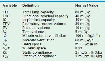
 of about 100 mL/kg/min in adults and 150 mL/kg/min in newborns. In healthy lungs, these settings should provide sufficient ventilation to maintain normal PaCO2 levels of approximately 40 mmHg, and should generate peak inspiratory pressures between 15–20 cmH2O above an applied PEEP of 5 cmH2O. Clinical assessment by observing chest wall movement, auscultation, and evaluation of gas exchange determines the appropriate Vt in a given patient.
 ), and the oxygen index (OI).


