Chapter 238 Measles
Measles is highly contagious and was once an inevitable experience during childhood. Owing to widespread vaccination, endemic transmission has been interrupted in the USA; indigenous or imported cases (in children or adults) have occasionally resulted in epidemics in the USA in unimmunized or partially immunized American or foreign-born children. In some areas of the world, measles remains a serious threat to children.
Etiology
Measles virus is a single-stranded, lipid-enveloped RNA virus in the family Paramyxoviridae and genus Morbillivirus. Other members of the genus Morbillivirus affect a variety of mammals, such as rinderpest virus in cattle and distemper virus in dogs, but humans are the only host of measles virus. Of the 6 major structural proteins of measles virus, the 2 most important in terms of induction of immunity are the hemagglutinin (H) protein and the fusion (F) protein. The neutralizing antibodies are directed against the H protein, and antibodies to the F protein limit proliferation of the virus during infection. Small variations in genetic composition have also been identified that result in no effect on protective immunity but provide molecular markers that can distinguish between viral types. These markers have been useful in the evaluation of endemic spread of measles.
Epidemiology
The measles vaccine has changed the epidemiology of measles dramatically. Once worldwide in distribution, endemic transmission of measles has been interrupted in many countries where there is widespread vaccine coverage. Historically, measles caused universal infection in childhood in the USA, with 90% of children acquiring the infection before 15 yr of age. Morbidity and mortality associated with measles decreased prior to the introduction of the vaccine as a result of improvements in health care and nutrition. However, the incidence declined dramatically following the introduction of the measles vaccine in 1963. The attack rate fell from 313 cases/100,000 population in 1956-1960 to 1.3 cases/100,000 in 1982-1988.
A nationwide indigenous measles outbreak occurred in 1989-1991, resulting in >55,000 cases, 11,000 hospitalizations, and 123 deaths, demonstrating that the infection had not yet been conquered. This resurgence was attributed to vaccine failure in a small number of school-aged children, low coverage of preschool-aged children, and more rapid waning of maternal antibodies in infants born to mothers who had never experienced wild-type measles infection. Implementation of the 2-dose vaccine policy and more intensive immunization strategies resulted in interruption of endemic transmission in the USA in 1993. The current rate is <1 case/1,000,000 population.
Measles continues to be imported into the USA from abroad; therefore, continued maintenance of >90% immunity through vaccination is necessary to prevent widespread outbreaks from occurring (Fig. 238-1).

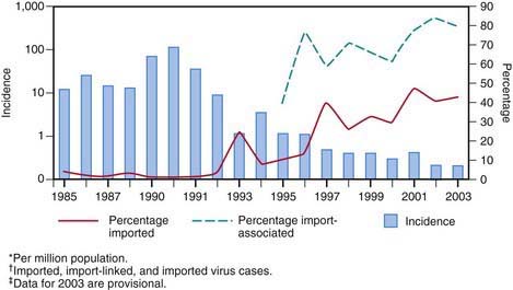
Figure 238-1 Incidence* and percentage of import-associated† measles cases, by year in the USA, 1985–2003‡.
(From the Centers for Disease Control and Prevention: Epidemiology of measles—United States, 2001–2003, MMWR Morb Mortal Wkly Rep 53:713–716, 2004.)
In 2008, 131 cases of measles were reported to the U.S. Centers for Disease Control and Prevention (CDC) in the first 7 months of the year, the highest year-to-date number of cases since 1996. Seven outbreaks accounted for 106 (81%) of the cases. Of the total, 17 patients (13%) had acquired the infection abroad and 99 cases (76%) were linked epidemiologically with the importations. These importation-associated cases occurred primarily among unvaccinated school-aged children whose parents had religious or philosophical objections to vaccination. Measles continues to be imported from abroad, and outbreaks will continue to occur in communities with low vaccination rates.
Transmission
The portal of entry of measles virus is through the respiratory tract or conjunctivae following contact with large droplets or small-droplet aerosols in which the virus is suspended. Patients are infectious from 3 days before to up to 4-6 days after the onset of rash. Approximately 90% of exposed susceptible individuals experience measles. Face-to-face contact is not necessary, because viable virus may be suspended in air for as long as 1 hr after the patient with the source case leaves a room. Secondary cases due to spread of aerosolized virus have been reported in physicians’ offices and in hospitals.
Pathology
Measles infection causes necrosis of the respiratory tract epithelium and an accompanying lymphocytic infiltrate. Measles produces a small vessel vasculitis on the skin and on the oral mucous membranes. Histology of the rash and exanthem reveals intracellular edema and dyskeratosis associated with formation of epidermal syncytial giant cells with up to 26 nuclei. Viral particles have been identified within these giant cells. In lymphoreticular tissue, lymphoid hyperplasia is prominent. Fusion of infected cells results in multinucleated giant cells, the Warthin-Finkeldey giant cells that are pathognomonic for measles, with up to 100 nuclei and intracytoplasmic and intranuclear inclusions.
Pathogenesis
Measles consists of 4 phases: incubation period, prodromal illness, exanthematous phase, and recovery. During incubation, measles virus migrates to regional lymph nodes. A primary viremia ensues that disseminates the virus to the reticuloendothelial system. A secondary viremia spreads virus to body surfaces. The prodromal illness begins after the secondary viremia and is associated with epithelial necrosis and giant cell formation in body tissues. Cells are killed by cell-to-cell plasma membrane fusion associated with viral replication that occurs in many body tissues, including cells of the central nervous system (CNS). Virus shedding begins in the prodromal phase. With onset of the rash, antibody production begins, and viral replication and symptoms begin to subside. Measles virus also infects CD4+ T cells, resulting in suppression of the Th1 immune response and a multitude of other immunosuppressive effects.
Clinical Manifestations
Measles is a serious infection characterized by high fever, an enanthem, cough, coryza, conjunctivitis, and a prominent exanthem. After an incubation period of 8-12 days, the prodromal phase begins with a mild fever followed by the onset of conjunctivitis with photophobia, coryza, a prominent cough, and increasing fever. Koplik spots represent the enanthem and are the pathognomonic sign of measles, appearing 1 to 4 days prior to the onset of the rash (Fig. 238-2). They first appear as discrete red lesions with bluish white spots in the center on the inner aspects of the cheeks at the level of the premolars. They may spread to involve the lips, hard palate, and gingiva. They also may occur in conjunctival folds and in the vaginal mucosa. Koplik spots have been reported in 50-70% of measles cases but probably occur in the great majority.

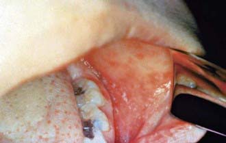
Figure 238-2 Koplik spots on the buccal mucosa during the 3rd day of rash.
(From Centers for Disease Control and Prevention: Public health image library, image #4500 [website]. http://phil.cdc.gov/phil/details.asp.)
Symptoms increase in intensity for 2-4 days until the 1st day of the rash. The rash begins on the forehead (around the hairline), behind the ears, and on the upper neck as a red maculopapular eruption. It then spreads downward to the torso and extremities, reaching the palms and soles in up to 50% of cases. The exanthem frequently becomes confluent on the face and upper trunk (Fig. 238-3).

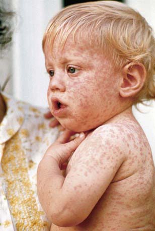
Figure 238-3 A child with measles displaying the characteristic red blotchy pattern on his face and body.
(From Kremer JR, Muller CP: Measles in Europe—there is room for improvement, Lancet 373:356–358, 2009.)
With the onset of the rash, symptoms begin to subside. The rash fades over about 7 days in the same progression as it evolved, often leaving a fine desquamation of skin in its wake. Of the major symptoms of measles, the cough lasts the longest, often up to 10 days. In more severe cases, generalized lymphadenopathy may be present, with cervical and occipital lymph nodes especially prominent.
Inapparent Measles Infection
In individuals with passively acquired antibody, such as infants and recipients of blood products, a subclinical form of measles may occur. The rash may be indistinct, brief, or, rarely, entirely absent. Likewise, some individuals who have received vaccine, when exposed to measles, may have a rash but few other symptoms. Persons with inapparent or subclinical measles do not shed measles virus and do not transmit infection to household contacts.
Children who had received the original formalin-inactivated measles vaccine at times demonstrated a more severe form of disease called atypical measles. Such patients had onset of high fever and headache followed by the appearance of a maculopapular rash on the extremities that become petechial and purpuric and progressed in a centripetal direction. The illness was frequently complicated by pneumonia and pleural effusions. It is thought that atypical measles was caused by development of circulating immune complexes that formed as a result of an abnormal immune response to the vaccine.
Laboratory Findings
The diagnosis of measles is almost always based on clinical and epidemiologic findings. Laboratory findings in the acute phase include reduction in the total white blood cell count, with lymphocytes decreased more than neutrophils. Absolute neutropenia has been known to occur, however. In measles not complicated by bacterial infection, the erythrocyte sedimentation rate and C-reactive protein level are normal.
Diagnosis
In the absence of a recognized measles outbreak, confirmation of the clinical diagnosis is often recommended. Serologic confirmation is most conveniently made by identification of immunoglobulin M (IgM) antibody in serum. IgM antibody appears 1-2 days after the onset of the rash and remains detectable for about 1 mo. If a serum specimen is collected <72 hr after onset of rash and is negative for measles antibody, a second specimen should be obtained. Serologic confirmation may also be made by demonstration of a fourfold rise in IgG antibodies in acute and convalescent specimens collected 2-4 wk later. Viral isolation from blood, urine, or respiratory secretions can be accomplished by culture at the CDC or local or state laboratories. Molecular detection by polymerase chain reaction (PCR) is possible but is a research tool.
Differential Diagnosis
Typical measles is unlikely to be confused with other illnesses, especially if Koplik spots are observed. Measles in the later stages or inapparent or subclinical infections may be confused with a number of other exanthematous immune-mediated illnesses and infections, including rubella, adenoviruses, enteroviruses, and Epstein-Barr virus. Exanthem subitum (in infants) and erythema infectiosum (in older children) may also be confused with measles. Mycoplasma pneumoniae and group A streptococcus may also produce rashes similar to that of measles. Kawasaki syndrome can cause many of the same findings as measles but lacks discrete intraoral lesions (Koplik spots) and a severe prodromal cough, and typically leads to elevations of neutrophils and acute-phase reactants. In addition, the characteristic thrombocytosis of Kawasaki syndrome is absent in measles (Chapter 160). Drug eruptions may occasionally be mistaken for measles.

Full access? Get Clinical Tree

