Diphtheria (Corynebacterium diphtheriae)

Chapter 180 Diphtheria (Corynebacterium diphtheriae)


Diphtheria is an acute toxic infection caused by Corynebacterium species, typically Corynebacterium diphtheriae and rarely toxigenic strains of Corynebacterium ulcerans. Although diphtheria was reduced from a major cause of childhood death to a medical rarity in the Western hemisphere in the early 20th century, current reminders of the fragility of this success emphasize the necessity to continue vigorous promotion of those same principles across the global community.


Etiology


Corynebacteria are aerobic, nonencapsulated, non–spore-forming, mostly nonmotile, pleomorphic, gram-positive bacilli. C. diphtheriae is by far the most commonly isolated agent of diphtheria. C. ulcerans is more commonly isolated from cattle and can cause similar disease. As corynebacteria are not fastidious in growth requirements, their isolation is enhanced by use of a selective medium (i.e., cystine-tellurite blood agar or Tinsdale agar) that inhibits growth of competing organisms and, when reduced by C. diphtheriae, renders colonies gray-black. Differentiation of C. diphtheriae from C. ulcerans is based on urease activity, because C. ulcerans is urease-positive.


Four C. diphtheriae biotypes (mitis, intermedius, belfanti, gravis) are capable of causing diphtheria and are differentiated by colonial morphology, hemolysis, and fermentation reactions. The ability to produce diphtheritic toxin results from acquisition of a lysogenic Corynebacteriophage by either C. diphtheriae or C. ulcerans, which encodes the diphtheritic toxin gene and confers diphtheria-producing potential on these strains. Thus, indigenous nontoxigenic C. diphtheriae can be rendered toxigenic and disease-producing after importation of a toxigenic C. diphtheriae and transmission of the bacteriophage. Demonstration of diphtheritic toxin production or potential for toxin production by an isolate is necessary to confirm disease. The former is done in vitro using the agar immunoprecipitin technique (Elek test) or in vivo with the toxin neutralization test in guinea pigs, the latter by polymerase chain reaction testing for carriage of the toxin gene. Toxigenic and nontoxigenic strains are indistinguishable by colony type, microscopic features, or biochemical test results.


Epidemiology


Unlike other diphtheroids (coryneform bacteria), which are ubiquitous in nature, C. diphtheriae is an exclusive inhabitant of human mucous membranes and skin. Spread is primarily by airborne respiratory droplets, direct contact with respiratory secretions of symptomatic individuals, or exudate from infected skin lesions. Asymptomatic respiratory tract carriage is important in transmission. Where diphtheria is endemic, 3-5% of healthy individuals can carry toxigenic organisms, but carriage is exceedingly rare if diphtheria is rare. Skin infection and skin carriage are silent reservoirs of C. diphtheriae, and organisms can remain viable in dust or on fomites for up to 6 mo. Transmission through contaminated milk and an infected food handler has been proven or suspected.


In the 1920s, >125,000 diphtheria cases, with 10,000 deaths, were reported annually in the USA, with the highest fatality rates among the very young and the elderly. The incidence then began to decrease and, with widespread use of diphtheria toxoid in the USA after World War II, declined steadily through the late 1970s. Since then, ≤5 cases have occurred annually in the USA, with no epidemics of respiratory tract diphtheria. Similar decreases occurred in Europe. Despite the worldwide decrease in disease incidence, diphtheria remains endemic in many developing countries with poor immunization rates against diphtheria.


When diphtheria was endemic, it primarily affected children <15 yr of age. Since the introduction of toxoid immunization, the disease has shifted to adults who lack natural exposure to toxigenic C. diphtheriae in the vaccine era and have low rates of booster immunization. In the 27 sporadic cases of respiratory tract diphtheria reported in the USA in the 1980s, 70% occurred among persons >25 yr of age. The largest outbreak of diphtheria in the developed world since the 1960s occurred from 1990 to 1996 in the newly independent countries of the former Soviet Union, involving >150,000 cases in 14 of 15 countries. Of these, >60% of cases occurred in individuals >14 yr of age. Case fatality rates ranged from 3% to 23% by country. Factors contributing to the epidemic included a large population of underimmunized adults, decreased childhood immunization rates, population migration, crowding, and failure to respond aggressively during early phases of the epidemic. Cases of diphtheria among travelers from these endemic areas were transported to many countries in Europe.


Most proven cases of respiratory tract diphtheria in the USA in the 1990s were associated with importation of toxigenic C. diphtheriae, although clonally related toxigenic C. diphtheriae has persisted in this country and Canada for at least 25 yr.


Cutaneous diphtheria, a curiosity when diphtheria was common, accounted for >50% of reported C. diphtheriae isolates in the USA by 1975. This indolent local infection, compared with mucosal infection, is associated with more prolonged bacterial shedding, greater contamination of the environment, and increased transmission to the pharynx and skin of close contacts. Outbreaks are associated with homelessness, crowding, poverty, alcoholism, poor hygiene, contaminated fomites, underlying dermatosis, and introduction of new strains from exogenous sources. It is no longer a tropical or subtropical disease; 1,100 C. diphtheriae infections were documented in a neighborhood in Seattle (site of the last major U.S. outbreak), from 1971 to 1982; 86% were cutaneous, and 40% involved toxigenic strains. Cutaneous diphtheria is an important source for toxigenic C. diphtheriae in the USA, and its importation is frequently the source for subsequent sporadic cases of respiratory tract diphtheria. To focus attention on respiratory tract diphtheria, the condition more likely to cause acute respiratory complications and toxic manifestations, C. diphtheria isolates from cutaneous disease were removed from annual diphtheria statistics reported by the Centers for Disease Control and Prevention (CDC) after 1979.


Clinical Manifestations


The manifestations of C. diphtheriae infection are influenced by the anatomic site of infection, the immune status of the host, and the production and systemic distribution of toxin.


Respiratory Tract Diphtheria


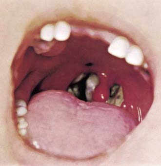
In a classic description of 1,400 cases of diphtheria in California (1954), the primary focus of infection was the tonsils or pharynx (94%), with the nose and larynx the next 2 most common sites. After an average incubation period of 2-4 days, local signs and symptoms of inflammation develop. Infection of the anterior nares is more common among infants and causes serosanguineous, purulent, erosive rhinitis with membrane formation. Shallow ulceration of the external nares and upper lip is characteristic. In tonsillar and pharyngeal diphtheria, sore throat is the universal early symptom: Only half of patients have fever, and fewer have dysphagia, hoarseness, malaise, or headache. Mild pharyngeal injection is followed by unilateral or bilateral tonsillar membrane formation, which can extend to involve the uvula (which may cause toxin-mediated paralysis), soft palate, posterior oropharynx, hypopharynx, or glottic areas (Fig. 180-1). Underlying soft tissue edema and enlarged lymph nodes can cause a bull-neck appearance. The degree of local extension correlates directly with profound prostration, bull-neck appearance, and fatality due to airway compromise or toxin-mediated complications (Fig. 180-2).


Stay updated, free articles. Join our Telegram channel

Jun 18, 2016 | Posted by in PEDIATRICS | Comments Off on Diphtheria (Corynebacterium diphtheriae)

Full access? Get Clinical Tree

Get Clinical Tree app for offline access