Fig. 8.1
Normal uterus; sagittal T2-weighted MRI clearly depicting the zonal anatomy of the uterus with the three zones displayed: intermediate signal of the endometrial lining (straight arrow), low signal of the junctional zone, i.e., the inner myometrium (curved arrow), and intermediate signal of the outer myometrium
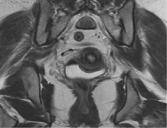
Fig. 8.2
Axial T2-weighted MRI showing three distinct zones of the uterus
The central endometrium has a high T2 signal due to the endometrium and secretions. The endometrium varies in thickness with the menstrual cycle and menopausal status. It can measure up to 14 mm in the secretory phase but is thinned in the follicular phase. Postmenopausal women should have a homogenous endometrium with a width less than 5 mm [3].
The myometrium is separated into (1) the inner myometrium, also known as the junctional zone which appears like a low-signal band on T2-weighted images, and (2) the outer myometrium which has an intermediate T2 signal.
When using oral contraceptives, the endometrium becomes thinned and the junctional zone less prominent.
After menopause, the junctional zone is thinned and not visualized consistently.
Post-contrast, the junctional zone shows the earliest enhancement, the outer myometrium enhances slightly later, and the endometrium enhances last.
The cervix shows four distinct zones on T2-weighted images:
1.
Central hyperintense-signal mucus.
2.
High-signal endocervical mucosa and glands.
3.
Hypointense fibrous stroma; this is continuous with the junctional zone of the uterus.
4.
Outer intermediate-signal loose stroma.
MRI in Endometrial Carcinoma
MRI is essential for preoperative staging because it demonstrates the depth of myometrial invasion which is the most important morphologic prognostic factor. The depth of myometrial invasion and histological grade correlate strongly with the presence of lymph node metastases and patient survival. Lymph node metastases prevalence increases from 3 % with superficial myometrial invasion to 46 % with deep myometrial invasion [4, 5].
Evaluation of the extent of myometrial invasion by gross inspection at surgery or at frozen section remains inaccurate in a significant number of patients [6]. Though the majority of patients present with Stage 1A where the standard treatment is total abdominal hysterectomy with bilateral salpingo-oophorectomy, the challenge is to identify patients at risk for recurrence who would require more radical surgery or adjuvant therapy and avoid overtreating low-risk patients [6]. Preoperative MRI hence helps in this decision making between lymph node sampling for Stage 1A disease and radical lymph node resection for Stage 1B disease.
MRI also assesses more advanced disease such as cervical stromal involvement. Gross cervical invasion could require preoperative radiation therapy or a different surgical approach such as a radical hysterectomy rather than a total abdominal hysterectomy. Adnexal involvement, uterine tumor size/volume, and the presence of ascites or nodal disease can also be assessed. These can help determine the surgical approach such as transabdominal, transvaginal, or laparoscopic.
Full assessment of the abdomen can be done to assess for lymph nodal involvement/ hepatic or peritoneal disease. In high-risk patients for surgery due to comorbidities, MRI is helpful in planning alternative therapy such as radiation or hormonal therapy for Stage 1 disease. Depth of myometrial invasion which is the most important morphologic prognostic factor can only be evaluated with MRI, as MRI is the only modality which demonstrates the zonal anatomy of the uterus.
The FIGO staging system was updated in 2009 with three important changes which are relevant to MRI.
Tumors confined to the endometrium (previous Stage 1A) and those involving the inner half of the myometrium (previous Stage 1B) are now combined together in Stage 1A. This actually improves the accuracy of MRI, as with the old system distinguishing between the two was difficult in some patients due to thinning/loss of the junctional zone or poor tumor to myometrium contrast.
One of the important changes in the FIGO staging system (2009) is the clubbing together of tumors confined to the endometrium (previous Stage 1A) and those involving the inner half of the myometrium (previous Stage 1B); both come under Stage 1A. This improves the accuracy of MRI (Figs. 8.3 and 8.4).
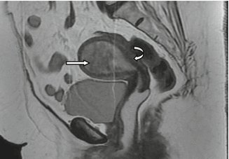
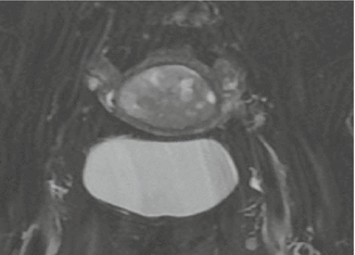

Fig. 8.3
81-year-old lady with postmenopausal bleed. Sagittal T2-weighted MRI showing a large mass filling the endometrial cavity (straight arrow). Intact junctional zone clearly demonstrated (curved arrow) with no extension of mass into the myometrium. No extension to the vagina

Fig. 8.4
Axial T2-weighted MRI showing the large mass filling the endometrial cavity. Intact junctional zone clearly demonstrated with no extension of mass into the myometrium
The other change is in Stage II. IIA tumors were those with endocervical glandular invasion, and IIB tumors were those with cervical stromal invasion. In the new system, endocervical glandular invasion is included in Stage 1 disease and those with cervical stromal invasion, Stage II disease.
Stage IIIA disease invades the serosa or adnexa (Figs. 8.5 and 8.6).
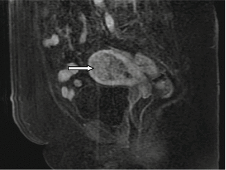
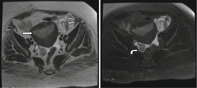

Fig. 8.5
Post-contrast sagittal T1 fat-suppressed image showing the mass in the endometrial cavity (straight arrow), enhancing less than the adjacent myometrium

Fig. 8.6
39-year-old lady with biopsy-proven endometrial carcinoma, axial T2 and T2 fat-suppressed images showing mass in the endometrial cavity (straight arrows) and extension to the bilateral fallopian tubes (curved arrows) – Stage IIIA disease
Stage IIIB disease involves the vagina or parametrium. Vaginal invasion is shown as segmental loss of the low-signal line of the vaginal wall. Parametrial involvement appears as disruption of the serosa with direct extension into the surrounding parametrium.
MRI Appearances
Endometrial carcinoma shows heterogeneous intermediate-signal intensity on T2-weighted images when compared to the normal hyperintense endometrium.
It is mildly hyperintense on T2-weighted images when compared to the myometrium. The depth of myometrial penetration with extension beyond/breach of the junctional zone is well seen on the high-resolution T2 images in the sagittal plane and axial oblique plane obtained perpendicular to the endometrial cavity except where there is thinning of the junctional zone or poor tumor to myometrium contrast.
In a postmenopausal woman, in addition, there is often an overall thinning of the myometrium due to uterine involution which can render accurate assessment of myometrial invasion difficult.
Other pitfalls which make accurate assessment difficult include extension into the cornua, compression of the myometrium by a large polypoid tumor/tumor filling the endometrial cavity with compression of the overlying myometrium, and presence of leiomyomas or adenomyosis.
In these cases, additional imaging such as dynamic contrast or diffusion-weighted imaging is of help. The depth of myometrial penetration with extension beyond/breach of the junctional zone is well seen on the high-resolution T2 images in the sagittal plane and most importantly in the axial oblique plane which is obtained perpendicular to the endometrial cavity.
Dynamic Contrast-Enhanced MRI
This has been shown to improve the diagnostic accuracy of MRI from 55 % to 77 % for routine non-contrast MRI to 85–91 % for dynamic contrast-enhanced images [6].
Stay updated, free articles. Join our Telegram channel

Full access? Get Clinical Tree


