Fig. 1
Typical appearance of a patient with Madelung deformity. Note the obvious appearance of a prominent ulnar with volar subluxation of the hand/carpus
There is some debate regarding the genetic component of Madelung’s deformity, although many cases are thought to be autosomal dominant with incomplete penetrance (Grigelioniene et al. 2001; Hirschfeldova et al. 2012; Benito-Sanz et al. 2005; Rappold et al. 2002). Leri-Weill syndrome (dyschondrosteosis ) is characterized by mesomelic dwarfism , short stature, and Madelung’s deformity (Fig. 2). These patients have been shown to have abnormalities in the short stature homeobox gene (SHOX) that resides in the pseudoautosomal region of the sex chromosomes. While the SHOX haploinsufficiency was originally thought to be solely caused by gene deletion, more recently investigators have also found it to be the result of mutations within the SHOX gene itself. Madelung’s deformity can also be seen as a component in Turner’s syndrome, Langer-Giedion syndrome, multiple hereditary exostoses, and multiple enchodromatoses. In addition, there are also patients of normal stature who have no other findings suggestive of underlying syndromes, but who have typical Madelung’s deformity.
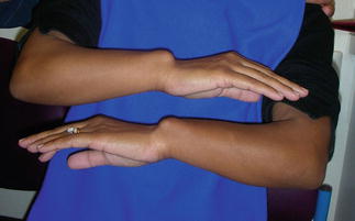

Fig. 2
A patient with Leri-Weill syndrome. Note the short forearms in association with the wrist findings
Pathoanatomy and Applied Anatomy
The patient with Madelung’s deformity has a characteristic position of the upper extremity. In most cases, the ulna appears prominent with the carpus and hand volarly translated. Pain over the ulna may be present with some decrease in forearm supination and wrist extension. The forearms themselves may be short. Plain radiographs can show a spectrum of deformity ranging from minimal increase in radial inclination to substantially increased radial inclination along with lunate subsidence (Fig. 3a, b). McCarroll et al. (2010) reported that radiographic criteria for the diagnosis included ulnar tilt of 33° or greater, lunate subsidence of 4 mm or more, lunate fossa angle of 40° or greater, and palmar carpal displacement of 20 mm or more. In the skeletally immature patient, premature closure of the volar-ulnar radial physis is noted. Radiographs of the entire forearm to include the elbows are important as whole-bone involvement of the radius has been documented and has implications on eventual outcomes (Zebala et al. 2007; Fig. 4). CT and MRI imaging can add a three-dimensional view of the deformity, but is usually not necessary to establish or treat the Madelung’s deformity patient.
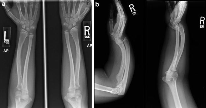
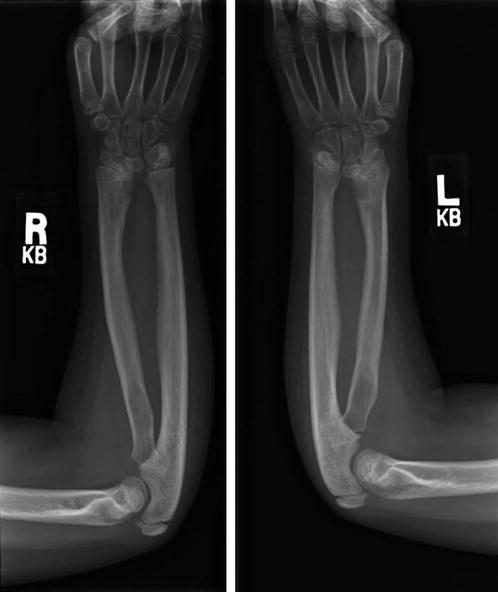

Fig. 3
(a) Radiographs of a patient with minimal deformity. (b) Radiographs of a patient with severe deformity

Fig. 4
A patient with whole-bone involvement of the radius
Vickers and Nielsen (1992) described a thick fibrous structure (the so-called Vickers’ ligament ), which begins on the ulnovolar metaphyseal region of the radius and attaches to the lunate and triangular fibrocartilage. Whether this anomalous structure is truly an additional structure or just prominence of the volar wrist ligamentous structures is subject of debate, but its presence has been reported in up to 91 % of patients (Vickers and Nielsen 1992; Nielsen 1977). Some authors believe that release of this structure during operative intervention is an integral part of the surgery (Vickers and Nielsen 1992; Nielsen 1977; Harley et al. 2006; Steinman et al. 2013). However, it is doubtful that the potential tethering effect of the “ligament” is the primary cause of deformity. Madelung’s deformity is not seen in the child less than 3 years of age (a period where rapid growth takes place).
Madelung’s Deformity Treatment Options
Nonoperative Treatment
Many patients display asymmetric involvement. Thus, even though one extremity warrants treatment because of symptoms, observation in the less involved extremity is indicated. In some patients, the physeal involvement on the volar-ulnar portion of the distal radius is minimal, with minimal deformity of the radius and carpus. Many of these patients, in fact, can be carefully evaluated every 6 months during their adolescent growth period until skeletally mature and may never need surgical intervention (Fig. 5a, b, c). Prophylactic release of Vickers’ ligament has not been proven to prevent deformity or pain in these patients.
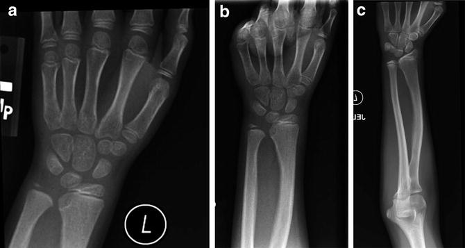

Fig. 5
Serial radiographs of a patient showing no progression of deformity over time
Madelung’s deformity | |
|---|---|
Nonoperative treatment | |
Indications | Contraindications |
Minimal deformity | Progressive deformity |
No pain | Pain |
Operative Treatment
Symptoms that warrant consideration for surgery in most patients include pain and unacceptable deformity. Early reports on operative techniques centered solely on the ulna with varying success (Ranawat et al. 1975; Glard et al. 2007; Bruno et al. 2003). Most surgical treatment now more logically addresses the radius as the deformity appears to be inherent to this bone (Vickers and Nielsen 1992; Harley et al. 2002, 2006; Steinman et al. 2013; Murphy et al. 1996). Age plays an important role with respect to which procedure is chosen. Vickers and Nielsen (1992) have reported excellent success with ligament release and physiolysis in those skeletally immature patients who have considerable growth potential remaining. In the patient with substantial deformity and not much forearm growth remaining, corrective radial osteotomy with ligament release is warranted.
Physiolysis and Ligament Release
After induction of general anesthesia and under tourniquet control, a longitudinal incision is placed over the flexor carpi radialis tendon (FCR) overlying the distal radius. Next, dissection is carried down to the pronator quadratus where the distal portion is divided to allow exposure of the abnormal radio-lunate ligament (Vickers’). It is easily identified in a concavity in the metaphyseal portion of the radius. This is carefully released from proximal to distal until the tethering of the lunate is released. This is usually readily apparent and results in being able to visualize the undersurface of the lunate. Next, physiolysis is performed as described initially by Langenskiold and later by Vickers (Vickers and Nielsen 1992). The down-turned metaphysis at the distal volar-ulnar radius is carefully trimmed to expose the remaining growth plate. Using a burr or rongeur, the abnormal physis is removed until more normal-appearing physeal cartilage is identified. Adequate resection is imperative to the success of this procedure. At this point, the area is then packed with a generous amount of autogenous fat that can be harvested from a number of donor sites (Fig. 6). It is important for this fat to make contact with the entire cavity that was created. Layered closure is performed and the extremity is immobilized for 3–4 weeks. After removal of the cast, range-of-motion (ROM) exercises are instituted and serial radiographs are performed to access growth and alignment (Fig. 7a, b, c).
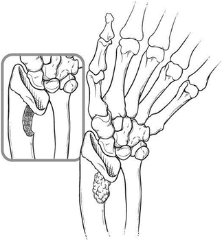
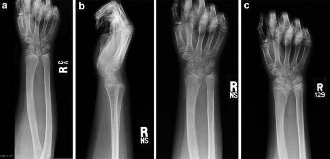

Fig. 6
A schematic drawing of the physiolysis/fat grafting procedure

Fig. 7
Serial radiographs of a patient who underwent physiolysis/fat grafting procedure
Physiolysis and ligament release for Madelung’s deformity |
|---|
Preoperative planning |
Patient in supine position with arm extended out on a hand table |
Use of C-arm brought in from end of hand table |
Sterile or non-sterile tourniquet |
Burr and rongeur |
Sterile prep of lower abdomen or similar area for autogenous fat graft harvest |
Physiolysis and ligament release for Madelung’s deformity |
|---|
Surgical steps |
Longitudinal incision overlying FCR in distal forearm |
Exposure and division of distal portion of pronator quadratus muscle, exposing Vickers’ ligament
Stay updated, free articles. Join our Telegram channel
Full access? Get Clinical Tree
 Get Clinical Tree app for offline access

|