Author(s)
Publication
Country
Percent
Number of children with macrodactyly/number of children with abnormal extremities/number of children examined
Ogino et al.
JHS 1986
Japan
0.5 %
5/943
Leung et al.
JHS 1982
Hong Kong
0.5 %
2/326
Flatt
1994
United States
0.9 %
26/2,758
Giele et al.
JHS 2001
Australia
0.8 %
5/509/257,430
Pathoanatomy and Applied Anatomy Relating to Macrodactyly
In the extremity affected by macrodactyly, the radial side is more frequently affected than the ulnar side coinciding with the median nerve being involved 85 % of the time and the ulnar nerve only 15 % (Flatt 1994). The soft tissue involvement tends to be greater on the volar side of the hand as well, but the nail and dorsal skin may thicken. This imbalance may lead to hyperextension of the digit. For most types of macrodactyly, the enlargement is more pronounced distally. It is three times more common for the macrodactyly to affect multiple digits rather than a single digit alone. These affected digits are beside each other and most commonly are the index and middle finger (Wood 1993). There has yet to be a report of macrodactyly affecting two digits with a normal digit between the two enlarged digits. If left to their own devices, macrodactylous digits may begin to show signs of vascular insufficiency. The affected phalanges show thickening of their cortex and widening of their medullary canals as they fill with fatty marrow. Often, enlargement of the bony skeleton does not proceed symmetrically, leading to deviation of the digit either radially or more commonly, in the ulnar direction (Flatt 1994).
With regard to NTOM, the perineurium is the most abnormal component of the nerves affected. There is also an increase in the number of axons and the size of each individual axon (Rios et al. 2013).
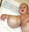
Other types of macrodactyly that are part of a syndrome have a broad spectrum of pathoanatomy . A multidisciplinary approach to these patients should be undertaken to not only care for their enlarged hand but also safely care for the entire child (Table 2).
Table 2
Comparison of overgrowth syndromes
Klippel-Trénaunay syndrome | Parkes Weber syndrome | Lipomatous overgrowth | CLOVE syndrome | Hemihypertrophy | Proteus syndrome | Maffucci syndrome | |
|---|---|---|---|---|---|---|---|
Capillary stain | Present, deep purple | Present, pink, often diffuse | Rare | Present, pink, diffuse, truncal | Uncommon | Present, pink, often diffuse | Absent (rarely reported) |
Progressive overgrowth | Present | Present | Present, symmetric | Present, often extensive | Either overgrowth or undergrowth | Absent at birth, haphazard, unrelenting, disproportional | Secondary to enchondroma growth, moderate to massive |
Hemodynamics | Slow-flow | Fast-flow | Slow-flow | Slow-flow | Slow-flow | Slow-flow | Slow-flow |
Vascular anomalies | VM, LVM | AVM, AVF | Absent | VM, LVM, LM | Absent | VM, LM, CM | VM within bone, hands common |
Associated anomalies | Common: GI, GU, genitalia | Rare, Cobb syndrome | Absent | No visceral | Skin, CNS, heart, GU, dental, others | Cerebriform soles/palms, linear nevi, dysregulated fat, lung cysts, facial neoplasms | None |
Limb enlargement | Moderate, disproportionate digits and toes common | Arm, leg length discrepancy | Moderate, nerve territory-oriented | Moderate | Macrodactyly, syndactyly, polydactyly, club feet | Major, asymmetrical | Disproportionate |
Limb affected | Upper 5 %, lower 95 % | Upper 23 %, lower 77 % | Equal | Equal | Arms, hands equally | Arms/legs, hands/feet equal | All |
Skeletal changes | Unusual, can occur with large lesions | Common, direct involvement and demineralization | Common, symmetric | Marked scoliosis, lower limb | Hip dysplasia, scoliosis, increased bone age | Skull hyperostosis, vertebral megaspondylodysplasia | Gross distortion, asymmetry, limb length discrepancy, scoliosis, short stature, fractures |
Macrodactyly | Diffuse enlargement | Minimal to moderate | Moderate to gigantic | Diffuse or longer with flexion contractures | Digits longer with flexion or extension contractures | Asymmetrical, moderate to gigantic | Digits longer with asymmetric masses, increased size due to tumors |
Dysregulated fat | Present within malformation | Present | Common, entire limb and axilla | Present | Multiple lipomas | Lipomas, regional fat absence | Present occasionally |
Nerve involvement | Absent | Absent | Intermittent | Intermittent | Absent, compression neuropathies with contractures | Absent | Absent |
Muscle anomalies | Displaced by VM, LVM | Displaced by malformation | Absent | Absent | Common, atavistic intrinsic and extrinsic muscles | Absent | None |
Associated neoplasms | Absent | Absent | Absent | Absent | Renal, adrenal and CNS | Ovarian, parotid | Chondrosarcoma (17.5–30 %), many others les frequently (ovarian, CNS) |
Coagulopathy | Present | Normal | Normal | Normal | Normal | Normal | None |
Cardiac failure | No | Yes | No | No | No | No | No |
Clinical prognosis | Stable | Often progressive deterioration, steal phenomenon. Prognosis is worse with CHF or amputation | Good, related to size and weight of limb | Progressive deterioration with extensive limb involvement | Progressive flexion, extension and intrinsic contractures | Progressive growth of affected regions | Good with local therapy; Ollier disease patients demonstrate increased problems |
Risks | DVT risk increased post op | Infection, CHF, gangrene | Secondary arthritis | Scoliosis, pulmonary compromise | Neoplasms (pheochromocytoma, Wilm’s tumor, hepatoblastoma) | Neoplasms, PE, depression | Pathologic fractures |
Genetics | Unknown | Unknown | Unknown | Unknown | Unknown | Unknown | Unknown |
— |  |  |  |  |  |  |  |
Assessment of Macrodactyly
With advancement in ultrasound techniques, macrodactyly can sometimes be diagnosed in utero (Yuksel et al. 2009), but typically is diagnosed at the time of birth by simple visualization and comparison to surrounding digits or contralateral unaffected digit. If the enlarged digit is not part of a syndrome, but is rather an isolated entity, the child must be monitored to see if the digit(s) grows in proportion to the child or not. Usually around the age of 2–3, the rate of growth, whether it is a static or progressive macrodactyly, will become evident by simple examination. The child should be followed at set intervals during these first couple of years determined by the anxiety of the parents, the magnitude of the enlargement, and the rate of growth.
The child should be watched playing and performing fine and gross motor activities with the arm and hand usually best accomplished with strategic bribery involving toys, stickers, and/or suckers. It should be noted whether the child uses the enlarged digit(s) or bypasses them entirely. During examination of the hand in these children, it is important to realize that overgrowth is not limited to a single digit or there is circumferential equal involvement even in a single digit. For example, the ulnar aspect of the index finger and radial portion of the middle finger may be enlarged and growing and deviating away from each other. This overgrowth will often extend into the palm and even up as high as the axilla. The fat distribution in the finger, palm, forearm, and arm is rarely circumferential but rather concentrated near the affected nerve and usually more severe distally. The shoulder, elbow, wrist, and all digits should be taken through a full active and passive range of motion to check for any loss of motion or scissoring of the digits.
The enlargement of the nerve and surrounding fat places the child at high risk for compressive neuropathies at the carpal, cubital, and/or radial tunnel. It is difficult to diagnose peripheral neuropathies in children. Wounds from chewing, biting, disuse, or ignoring the digit can all be signs that the child has lost sensation in the affected digit. The wrinkling test may prove useful if the child is amenable. The test requires warmwater submersion of the digit for 5 min to assess for skin wrinkling. Lack of wrinklings is indicative of loss of sympathetic innervation and nerve innervation. Nerve conduction studies and electromyography can be helpful to diagnosis and/or confirm peripheral neuropathies. Electrodiagnostic studies in children may require sedation, although oversedation will affect the study quality. Infants under 1 year of age can usually be tested without any sedation (or sedation only for the parents), while the 2- to 6-year-old children are the most likely to need mild sedation. A patient-savvy neurologist may succeed without pharmacologic aide by first doing the nerve conduction study and then electromyography using single fiber stimulation techniques (Hays et al. 1993).
Serial radiographs should be taken to monitor bony growth to help provide information concerning angular deformities. Serial studies also help guide the timing of epiphysiodesis. The same-sex parent should be examined and even radiographed if needed to provide a reference for the approximate final desired size of the digit for the child.
Magnetic resonance images can be useful for cases in which there is some component of vascular malformation in the affected limb.
When examining macrodactyly that is part of a syndrome such as Maffucci syndrome or neurofibromatosis, the physician must have a heightened awareness of the possibility of malignant transformation of a tumor of the hand or other body part. A substantial increase in pain may be the harbinger of the transformation of a plexiform neurofibroma to a neurofibrosarcoma, or a change in a skin lesion in Maffucci syndrome may signal the sarcomatous degeneration of a hemangioma or lymphangioma.
Other than grouping the types of overgrowth, currently there is no classification system that can guide treatment algorithms and compare treatment outcomes.
The three most accepted classification systems for overgrowth were published by De Laurenzi, Temtamy and McKusick, and Flatt, which divided digit overgrowth into groups based on growth rate, syndromal distinction, and disease process, respectively. Currently, Flatt’s classification with Upton’s modification is the most widely used when discussing macrodactyly.
Feriz, Werthemann, and Laurenzi recognized that not all large digits in the newborn grew at the same rate or for the same length of time. They were able to identify static macrodactyly or macrodactylia simplex congenital and differentiate it from the progressive macrodactyly or macrodystrophia lipomatosa progressiva (Feriz 1925; Werthemann 1952).
Temtamy and McKusick recognized that macrodactyly could be part of a syndrome or an isolated anomaly. They further subdivided isolated macrodactyly into true macrodactyly or pseudomacrodactyly. The authors declared that macrodactyly caused by hemangiomas, soft tissue tumor, edema from constriction band syndrome, arteriovenous aneurysms, or fistulas lead to soft tissue increase in size. However, these etiologies do not cause bone enlargement and therefore are not truly macrodactyly, hence the term pseudomacrodactyly. They recognized partial gigantism, neurofibromatosis, Ollier disease, Maffucci syndrome, Klippel-Trenaunay-Weber syndrome, and congenital lymphedema as all syndromes that could have macrodactyly as part of the spectrum of anomalies (Temtamy and McKusick 1978; Table 3).
Table 3
Classification of macrodactyly
Author (s) | Publication | Classification criteria |
|---|---|---|
De Laurenzi | G Med Milan 1962 | Growth rate |
Temtamy and McKusick | March of Dimes. Birth Defects 1978 | Syndromal or nonsyndromal |
Flatt | The Care of Congenital Hand Anomalies 1994 | Disease processes types I–III, type IV modification added by Upton (2005) |
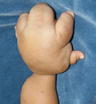
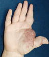
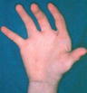
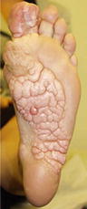
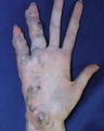
Flatt devised a classification system that divided macrodactyly into three types (Table 4). Type I is gigantism and lipofibromatosis. This type is equivalent to Kelikian’s nerve territory-oriented macrodactyly (NTOM). This group is further divided into static and progressive forms that were noted in earlier publications (Figs. 1 and 2).
Table 4


Flatt’s classification system of macrodactyly
Stay updated, free articles. Join our Telegram channel
Full access? Get Clinical Tree
