Lower respiratory tract infections and abnormalities

14.5 Lower respiratory tract infections and abnormalities


Lower respiratory tract infections


Lower respiratory tract infections (LRTIs) are a major cause of morbidity in developed and developing countries and the greatest cause of mortality in children under 5 years of age in developing countries, with more than 2 million deaths from pneumonia in this age group annually. Respiratory tract infections often involve both the upper and lower respiratory tracts, particularly those due to viruses. For respiratory tract infections that appear to involve mainly the upper airway, involvement of the lower respiratory tract is usually present to some degree. Over the last few years, acute asthma has been shown to be caused mainly by respiratory viral infection of the lower airway, and this is now recognized as the most common form of LRTI. However, this form of LRTI is discussed in the chapter on asthma (see Chapter 14.3).


Pneumonia


Pneumonia is a common cause of morbidity and mortality in children and is characterized by infection, inflammation and consolidation of the lung. There are many different causes of pneumonia. Viruses are the most common cause. Bacteria cause fewer cases of pneumonia, but the morbidity and mortality is several times higher than for viral pneumonia. Atypical infectious agents cause fewer cases of pneumonia, except in the increasing number of children with human immunodeficiency virus (HIV) infection.


Symptoms of acute infective pneumonia include dyspnoea, fever and malaise. Cough may be dry or moist, but is not always present. Pleuritic chest pain is often present. If the pneumonia involves the apices, neck pain may be present and can be confused with the neck stiffness of meningism. If the diaphragmatic pleural surface is involved, pain can be referred to the abdomen or shoulder tip.


Signs include tachypnoea and respiratory distress, dullness to percussion, and, on auscultation, localized crackles and bronchial breathing. Of these signs, tachypnoea is the most consistent and reliable, and may be the only sign pointing to this diagnosis. Pneumonia should be suspected in any child with an unexplained tachypnoea. Cyanosis is rare, but low levels of oxygen saturation are common. Clinical signs are less reliable in younger children and can be virtually absent in the presence of obvious radiological changes.


However, none of the symptoms or signs is specific for pneumonia, and the clinical diagnosis should be suspected when the history and examination are consistent. Signs of complications of pneumonia include those related to:


Investigations


Chest radiography is the most reliable investigation to confirm the presence of pneumonia. If the chest radiograph is normal, pneumonia can be reasonably excluded at that time, but if the X-ray is taken very early in the disease process this does not preclude radiological changes developing later. In general, patchy or peripheral consolidation may be more in keeping with a viral infection, lobar opacification is suggestive of bacterial pneumonia, and a more central peribronchial infiltrate may indicate Mycoplasma infection, but the specificity of these changes is relatively poor. Importantly, all of these radiological features can be found with asthma.


Repeat X-ray to establish resolution of the pneumonia is important to reduce the risk of missing an unrecognized, underlying or unresolved pathology, particularly those related to mechanical obstruction of the airway. Preferably this should be done at least 4–6 weeks after the acute illness has settled down, as the radiological abnormalities of pneumonia can be slow to resolve and may still be present if the repeat X-ray is done too early.


Blood culture may be performed if clinically indicated. Bacteraemia is not common in bacterial pneumonia.


A nasal aspirate should be taken if the diagnosis is unclear or viral aetiology is suspected. The aspirate should be subjected to polymerase chain reaction (PCR) analysis to detect the presence of causative respiratory viruses, but care is needed in interpreting results as positive results for respiratory viruses are common in asymptomatic children, and the presence of a virus in a nasal aspirate does not exclude the possibility of a bacterial pneumonia. Human rhinovirus detection should be included in the PCR detection panel as increasing recent evidence suggests that this virus is responsible for many LRTIs in hospitalized children. Rhinovirus typing may also be included in the near future, as even more recent evidence has shown that the newly discovered human rhinovirus group C is the most common virus group causing LRTI in hospitalized children, and also causes more pathology than previously known rhinovirus groups, as well as the majority of acute asthma admissions in children.


Sputum is often difficult to obtain and of limited usefulness due to contamination by upper airway bacteria.


Pleural fluid specimen. In more serious cases where a pleural effusion of sufficient size is present, obtaining pleural fluid should be considered, as it provides a more reliable possibility of obtaining a bacteriological diagnosis. Ultrasonography may be employed to guide the aspiration.


Bacterial antigen detection in the peripheral blood is also of limited use.


Immune function. In recurrent or atypical pneumonia, consideration should be given to the possibility of immunodeficiency. Initial investigations may include assessment of serum immunoglobulins and tests for HIV.


Pneumococcal pneumonia


Streptococcus pneumoniae is the most common cause of bacterial pneumonia in children at any age. Pneumococcal pneumonia is most common in children under 3 years of age. Risk factors include male sex, Indigenous ethnicity and pre-term delivery.


Pneumococcal pneumonia may be preceded by symptoms suggestive of a mild upper respiratory tract infection, and typical symptoms and signs of pneumonia may then appear. Although these can be non-specific, compared with viral pneumonia, symptoms are likely to include fever, dyspnoea, pleuritic chest pain and cough. Cough can, however, be absent and sputum production is less likely in younger children. Signs are more likely to include tachypnoea, grunting, nasal flaring, reduced movement of the chest wall on the affected side, dullness to percussion, reduced breath sounds and bronchial breathing over the area involved. Dullness to percussion may indicate the presence of an empyema. If the upper lobes are involved, neck stiffness may be present and lead to the misdiagnosis of meningitis.


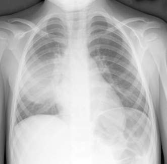
Chest X-ray findings vary widely, but the most common and classic finding is lobar involvement (Fig. 14.5.1). A well-defined round opacification is not uncommon and patchy changes can occur. Empyema, abscesses and pneumatoceles are less common than in staphylococcal pneumonia. Increases in white cell count and indices of inflammation are commonly found in peripheral blood, and blood culture may be positive.


The diagnosis should be made as early as possible and treatment commenced with penicillin and a third-generation cephalosporin. The response to treatment is usually rapid and complete recovery can be expected.


Staphylococcal pneumonia


Pneumonia due to Staphylococcus aureus is important as it is usually more severe than pneumonia due to other infective agents and is more common in younger children, especially those under 1–2 years of age. However, even in this younger age group, pneumococcal pneumonia is more common. Another important risk factor for staphylococcal pneumonia is a socially disadvantaged or Indigenous background.


Compared with other forms of pneumonia, the child with staphylococcal pneumonia is more likely to have a shorter acute history, to appear more unwell, and to have a high fever, marked tachypnoea and significant respiratory distress. The onset is usually acute and the course more rapid. Chest signs are often non-specific. The chest X-ray may be normal early in the disease, but later is more likely to show severe involvement. The early radiological features may be similar to those of other forms of bacterial pneumonia, including lobar consolidation, patchy shadowing and a small pleural effusion. However, more serious complications are common and these can develop quickly within the first few days; they include:


widespread opacifications, displaced intrathoracic structures and pleural effusions


more specific to staphylococcal pneumonia:


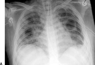
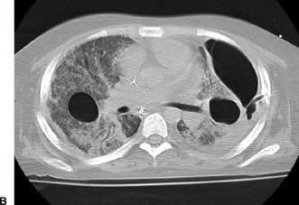
air leaks are common, occurring in around half of cases and include pneumothorax, pneumomediastinum, pneumopericardium and, in particular, pneumatoceles (Fig. 14.5.2A). Although highly specific to staphylococcal pneumonia, these complications are not pathognomonic of this condition, as air leaks including pneumatoceles can very occasionally be found in bacterial pneumonias caused by pneumococci, Escherichia coli, Klebsiella, Pseudomonas and group A streptococci.


High-resolution computed tomography (HRCT) of the chest (Fig. 14.5.2B) is often useful in defining the nature and extent of these complications.


Management


Owing to the increased risks from staphylococcal pneumonia, infants in whom this diagnosis is suspected should be hospitalized to allow adequate observation during the acute phase of the illness. Deterioration can be rapid, and air leaks can occur and require immediate treatment. Broad-spectrum antibiotics should be used until an accurate diagnosis can be made. The combination of a β-lactamase-resistant penicillin such as flucloxacillin and a third-generation cephalosporin, both given intravenously, is useful in this situation, as it combines direct treatment of staphylococci as well as coverage of other common respiratory pathogens. In any child under 2 years of age with clinically significant pneumonia, flucloxacillin should be included in the treatment regimen because of the much higher prevalence of staphylococcal pneumonia in this age group.


Resistance to β-lactamase-resistant penicillins (methicillin-resistant Staphylococcus aureus (MRSA)) is now common in staphylococcal pneumonia caused by community-acquired organisms, and multiresistant organisms have become more common in nosocomially acquired cases. Hence, from a therapeutic viewpoint, nosocomial staphylococcal infections are more likely to show multiple drug resistance than community-acquired MRSA infections. Treatment needs to be tailored to the prevailing local situation, but other drugs such as clindamycin should be considered. Given the high risk of relapse, the duration of antibiotic treatment is often extended to around 6 weeks, particularly in more severe cases or those with complications.


Stay updated, free articles. Join our Telegram channel

Aug 4, 2016 | Posted by in PEDIATRICS | Comments Off on Lower respiratory tract infections and abnormalities

Full access? Get Clinical Tree

Get Clinical Tree app for offline access