Liver diseases

20.5 Liver diseases


Normal liver anatomy and function


In evaluating liver disease, it is useful to think of the liver as being composed of two morphological and functional subunits. Injury to either compartment gives rise to a specific pattern of symptoms and liver test abnormalities. The first subunit is the parenchymal compartment consisting of hepatocytes, the carriers of anabolic and catabolic enzymes, and non-hepatocytic parenchymal cells. The latter include stellate cells, important for production of fibrous tissue under disease conditions, and the Kupffer cells, resident mononuclear leukocytes fulfilling a crucial role in processing antigens that enter the liver through the portal vein. The second subunit is the biliary compartment consisting of the canalicular and biliary system. This system carries bile produced by the hepatocytes into the bowel, but is also actively involved in resorption of various solutes. Bile acids, comprising 12% of bile, are required for the normal absorption of fat and fat-soluble vitamins in the intestinal lumen.


Blood supply to the liver is predominantly via the portal vein (70%), channelling intestinal nutrients towards the liver. The remaining 30% is provided by the hepatic artery, which supplies oxygen-rich blood to the parenchymal and biliary compartments. The bile ducts are sensitive to reduced arterial flow and may become damaged if hepatic arterial flow is interrupted. Blood flows out of the liver via the hepatic veins, which connect to the inferior vena cava.


The liver plays an indispensable role in whole-body energy homeostasis and provides the necessary enzymatic repertoire for numerous catabolic and anabolic pathways. The main functions of the liver are:


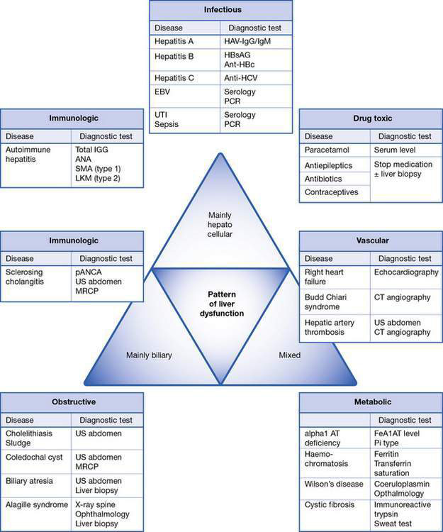
Pattern recognition in acute and chronic liver dysfunction


Assessment of liver dysfunction in the child is part of a diagnostic process involving a thorough clinical history, clinical symptoms and signs, and a complete panel of ‘liver function tests’ (see below). The integration of these results will allow further categorization of the disease process. Depending on which morphological and functional compartment is affected, liver disease can be primarily hepatocellular (parenchymal), biliary (cholestatic) or mixed hepatocellular–biliary, and can finally lead to failure of synthetic and detoxification function. Allocation of laboratory tests to a disease pattern results in more targeted investigations (Fig. 20.5.1).


Laboratory patterns of liver dysfunction


The actual term ‘liver function tests’ is a misnomer: not all measurements (e.g. alanine aminotransferase (ALT), aspartate aminotransferase (AST)) actually assess the function of the liver, whereas others (e.g. international normalized ratio (INR)) will not be obtained on request of ‘liver function tests’ on a biochemistry form. For practical purposes, the liver function tests can be divided into four categories, as described below.


Markers of biliary dysfunction/cholestasis: ALP, GGT, conjugated bilirubin, serum bile acids


Alkaline phosphatase (ALP) is an enzyme of uncertain physiological function. Blockage of bile flow leads to de novo production within the bile duct epithelium and to a rise in serum levels, which takes a few days. ALP levels may therefore be normal in acute biliary obstruction. The specificity of ALP is limited, as it is also found in other tissues including bone, intestine and kidney. ALP concentration is therefore often increased in growing children, particularly rapidly growing adolescents. If there is confusion, the origin of the ALP (i.e. liver or bone) can be tested in the laboratory by determining the so-called isoenzymes.


γ-Glutamyltransferase (GGT) is a more sensitive and specific test for biliary disease, and its level increases with biliary obstruction or inflammation. GGT is also induced by certain medications; therefore, if there is an isolated rise in GGT levels, take a full drug history. Bilirubin is the end-product of enzymatic haem breakdown, a component of haemoglobin. This unconjugated bilirubin is lipid-soluble, can cross membranes such as the blood–brain barrier and is therefore toxic, particularly in small infants. Conjugation of the bilirubin to glucuronide in the liver makes it water-soluble and allows excretion (via a transport protein in the cell membrane) into the small bile ducts, called canaliculi. The conjugated bilirubin then drains through the common bile duct into the duodenum. Damage to the transmembranous transport of conjugated bilirubin (e.g. hepatocellular insult) or blockage of the biliary drainage system (e.g. due to gallstones or biliary atresia) impairs bile flow, leading to a clinical phenotype called cholestasis. The water-soluble conjugated bilirubin flushes back into the bloodstream (conjugated hyperbilirubinaemia), is deposited in the skin (jaundice) and filtered in the kidneys (dark urine). The lack of bilirubin in the intestine results in pale (acholic) stools. Determination of conjugated bilirubin is the most important test in any jaundiced patient. A conjugated bilirubin fraction of more than 20% of total bilirubin is called conjugated hyperbilirubinaemia and indicates liver disease and impaired bile flow (cholestasis). Lack of bile flow also leads to retention and blood accumulation of bile acids and other components of bile. It is not known which of these components is the main mediator of the intense pruritus seen in chronic cholestasis.


Clinical signs of liver dysfunction


A thorough history and clinical examination are essential to guide the diagnostic approach in a child with suspected liver disease. When trying to identify the cause of liver disease, it is useful broadly to categorize children into infants versus older children, acute versus chronic liver disease, and biliary (cholestatic) versus hepatocellular injury. Of course, there will be significant overlap, but the categories are described in detail below and in Table 20.5.1 and Fig. 20.5.1.


Table 20.5.1 Clinical and aetiological categories of liver disease in childhood


Mode of presentation Time of presentation
Infancy Older child
Acute Infectious

Biliary/obstructive


Metabolic


Ischaemic


Immunological


Infectious

Biliary/obstructive


Metabolic


Drug-induced


Stay updated, free articles. Join our Telegram channel

Aug 4, 2016 | Posted by in PEDIATRICS | Comments Off on Liver diseases

Full access? Get Clinical Tree

Get Clinical Tree app for offline access