Case notes
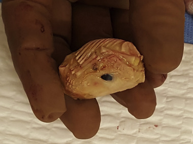
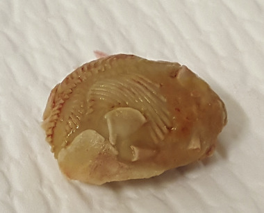
A 47-year-old woman came to a surgical mission in Haiti in 2016 desiring surgical management of heavy menstrual periods. She reported having had 5 pregnancies and deliveries. She denied any history of other medical problems or surgeries but had received little medical care in her life because of poor access to health care. An examination revealed a 16-week uterus consistent with fibroid tumors. Total abdominal hysterectomy, left salpingo-oophorectomy, and right salpingectomy were performed. On initial intraoperative examination of the pelvic cavity, significant adhesions were present that involved the uterus and bilateral adnexa; adhesions were filmy in nature but extensive. The right ovary was obscured by adhesions that encased it against the right pelvic sidewall, so it was left in situ. Although these findings suggested a history of pelvic inflammatory disease, shortly after, an alternative explanation presented itself when a lithopedion was discovered in the posterior cul-de-sac ( Figure 1 ). The lithopedion was free-floating and was removed without requiring disruption of adhesions or anatomy. The fetal skeleton was well-preserved with many identifiable structures that included humeri, radii, ulnas, metacarpals and phalanges, ribs, a scapula, and an eyeball. The gestational age was estimated at 13 weeks by a humerus length of 12 mm ( Figure 2 ).
Stay updated, free articles. Join our Telegram channel

Full access? Get Clinical Tree


