Linear Echoes in Amniotic Fluid
Anne Kennedy, MD
DIFFERENTIAL DIAGNOSIS
Common
Synechiae
Dichorionic Diamniotic Twins
Monochorionic Diamniotic Twins
Chorioamniotic Separation
Placental Abruption, Old
Uterine Septum
Placental Cysts
Less Common
Amniotic Band Syndrome
Circumvallate Placenta
ESSENTIAL INFORMATION
Key Differential Diagnosis Issues
Single or multiple gestation?
If multiple, inter-twin membrane is most likely cause of a linear echo in the amniotic fluid
Does linear echo cross cavity from side to side?
Placental edge to placental edge → circumvallate placenta
Uterine wall to uterine wall → synechia
Does linear echo parallel wall of uterine cavity?
How do linear echoes relate to placenta?
How do linear echoes relate to fetus?
Is there a history of intervention?
Prior uterine instrumentation
D&C, myomectomy, metroplasty
Procedure performed during current pregnancy?
Amniocentesis, amnioreduction, intrauterine transfusion, laser therapy or radiofrequency ablation
Helpful Clues for Common Diagnoses
Synechiae
Shelf or band-like structure which does not restrict fetal movement
Extra-amniotic: Fetal membranes wrap over synechiae
Straight, bulbous free edge with thinner sheet extending to endometrial surface
Y-shaped notch at endometrial base, created by membranes separating at endometrial margin
Placenta can abut or even wrap around synechia
Color Doppler may demonstrate flow within synechiae
In first trimester may cause distortion of gestational sac shape
May no longer be visible in 3rd trimester due to rupture or compression
Dichorionic Diamniotic Twins
Thick echogenic chorion completely surrounds each embryo
“Twin peak” sign: Wedge of chorionic tissue extending into base of inter-twin membrane
Two fetuses in separate chorionic sacs
Two amniotic sacs with two yolk sacs
Dichorionic membrane actually separates two amniotic cavities (i.e., extra-amniotic)
Monochorionic Diamniotic Twins
Two fetuses in single chorionic sac containing two amniotic sacs
Thin inter-twin membrane formed by two layers of amnion without interposed chorion
No “twin peak”
Twins must be same gender
Chorioamniotic Separation
Persistent unfused amnion and chorion > 16 wks
Amniotic membrane separate from uterine wall
Complete: Attached only at placental cord insertion site
Incomplete: Unattached around part of the uterine cavity, the commonest form
May be primary non-fusion
Look for signs of aneuploidy
May occur secondary to amniocentesis or fetal intervention
Increases risk of membrane rupture in twins → functional monoamniotic state → ↑ risk of cord entanglement
Placental Abruption, Old
Hypoechoic blood clot near or behind placenta
Marginal (most common), retroplacental or preplacental
Marginal: Bleed at edge of placental disc, dissects between chorion and uterine wall
Retroplacental does not cause confusion for linear echoes in amniotic fluid as located between placenta and myometrium
Preplacental abruption is rare
Hematoma on fetal surface of placenta
Clot may compress cord if close to insertion site
Subacute
May contain fluid-fluid level, septations common
Old
Liquefying blood, eventually sonolucent and may mimic amniotic fluid
Hemorrhage can dissect under chorionic membrane
Clot seen at a distance from placenta
Look in front of cervical os
Intraamniotic blood common → echogenic fluid → echogenic fetal bowel from swallowed blood
In twins, rarely, hematoma dissects between membranes
When old may appear as fluid-filled mass between membranes → increased linear interfaces in amniotic fluid
Uterine Septum
Midline, arising from fundus
In first trimester use 3D to create coronal images and confirm location, assess fundal contour
May be fibrous or composed of myometrium
Thicker than synechiae
Creates two distinct endometrial cavities
Placental Cysts
Chorionic cysts are simple cysts on fetal placental surface
Often near cord insertion site
If multiple may appear to create linear echoes in amniotic fluid
Curvilinear shape and relationship to placenta indicate etiology
If large, or if hemorrhage occurs, may compress cord
Helpful Clues for Less Common Diagnoses
Amniotic Band Syndrome
Entrapment of fetal parts by disrupted amnion
Amniotic band in contact with deformity, extends to uterine wall
Bands in amniotic fluid appear as multiple thin membranes
No flow in band on Doppler evaluation
Circumvallate Placenta
Placental margin elevated off uterine wall
Scanning parallel to edge → “marginal shelf”
Scanning longitudinally → “curled lip” of placental margin
Other Essential Information
Most linear echoes in amniotic fluid are of little clinical significance
Amniotic band syndrome can be lethal depending on extent of band-related damage
Image Gallery
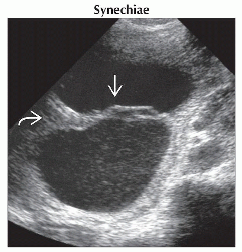 Ultrasound shows linear echoes
 crossing the amniotic cavity due to synechia. Note Y-shaped base crossing the amniotic cavity due to synechia. Note Y-shaped base  . The patient had a history of multiple D&C procedures for recurrent abortions. . The patient had a history of multiple D&C procedures for recurrent abortions.Stay updated, free articles. Join our Telegram channel
Full access? Get Clinical Tree
 Get Clinical Tree app for offline access

|