Large Bladder
Paula J. Woodward, MD
DIFFERENTIAL DIAGNOSIS
Common
Normal
Posterior Urethral Valves (PUV)
Less Common
Prune Belly Syndrome
First Trimester Megacystis
Rare but Important
Urethral Atresia
Megacystis Microcolon
ESSENTIAL INFORMATION
Helpful Clues for Common Diagnoses
Normal
Transient finding with otherwise normal urinary tract & amniotic fluid volume
Fetus will usually void during exam
Follow-up if bladder fails to decompress
Posterior Urethral Valves (PUV)
Urethral membrane acts as valve, resulting in bladder outlet obstruction
Occurs exclusively in males
“Keyhole” sign: Distended bladder “funnels” into dilated posterior urethra
Bladder often thick-walled, with degree of distention depending on severity of obstruction
Hydronephrosis common with potential development of renal dysplasia
Typically oligohydramnios, or even anhydramnios, in severe obstruction
Helpful Clues for Less Common Diagnoses
Prune Belly Syndrome
Triad of dramatic collecting system dilatation, deficiency of abdominal musculature & cryptorchidism
Often difficult to differentiate from PUV
Look carefully at urethra
Entire urethra may be dilated
Does not terminate at posterior urethra
May see spontaneous voiding
First Trimester Megacystis
Bladder length > 7 mm at 10-14 weeks
25% reported to have aneuploidy (trisomy 13, trisomy 18 most common)
Of those that are chromosomally normal, 90% regress while 10% progress to obstructive uropathy
Helpful Clues for Rare Diagnoses
Urethral Atresia
Complete obstruction, therefore massive bladder dilatation and anhydramnios
Occur in either males or females, but oligohydramnios often precludes ability to determine sex
Often indistinguishable from severe PUV
Megacystis Microcolon
Dilated bladder with normal to increased amniotic fluid
Differentiates it from other causes of large bladder
Intestinal hypoperistalsis may result in dilated small bowel
More common in females (M:F, 1:4)
Image Gallery
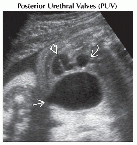 Coronal oblique ultrasound shows a dilated bladder “funneling” into a dilated posterior urethra
 (“keyhole” sign). There is also ureteral dilatation (“keyhole” sign). There is also ureteral dilatation  and hydronephrosis and hydronephrosis  . .Stay updated, free articles. Join our Telegram channel
Full access? Get Clinical Tree
 Get Clinical Tree app for offline access

|