Fig. 13.1
Lich-Gregoir procedure
A broad-spectrum antibiotic is routinely administered intravenously on induction of general anesthesia. A cystoscopy may be performed initially if bladder control is required, especially in children with a duplex system to assess the location of the ureteral orifices and to check the anatomy. Sometimes in these duplex systems, when an upper pole nephrectomy is scheduled during the same operation, a ureteral catheter can be placed in one of the two ureters to facilitate its recognition during the nephrectomy; otherwise, the ureterocele can be opened widely during the endoscopy before the upper pole nephrectomy and LER of the two ipsilateral ureters. In other circumstances, for example, bilateral reflux with ipsilateral grade I and contralateral grade III VUR, it is possible to do an endoscopic subureteric injection (STING procedure) to treat the grade I reflux before LER of the grade III reflux.
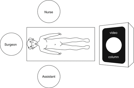
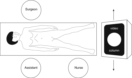
At the end of the cystoscopy, the child is placed in a supine position on the table with legs apart; the table is adapted to the size of the child to allow a good position of the video column, the closest as possible to the feet, to avoid a monitor too far from the surgeon (Fig. 13.2). After preparation of the abdominal wall, an urethral catheter is placed, and the bladder is emptied to allow a good vision of the pelvic cavity. The surgeon is positioned at the head of the patient; when the child is too old, he or she must to be placed laterally, on the right side for the left ureter and on the left side for the right ureter. The assistant and the nurse are placed according the position of the surgeon (Fig. 13.3).


Fig. 13.2
Position of the crew and equipment. The head of patient is close from the edge of the table. The surgeon, the telescope, the bladder, and the monitor form a straight line

Fig. 13.3
Position with a larger child
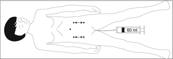
The laparoscopy is performed through a lateral or trans-umbilical incision under vision, and a 5 mm trocar is introduced for the telescope. The two 3 mm trocars are placed under direct vision at the left and right abdomen and at the same level from the umbilicus. When the child is small, the trocars are higher, and in adolescent they are in the lower part of the abdomen (Fig. 13.4).


Fig. 13.4
Position of trocars, more or less high according the size of patient. A sterile 60 ml syringe is set up into the bladder catheter to empty or fill the bladder during procedure
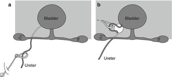
The first step is to release the ureter from the level of iliac vessels to the posterior wall of the bladder. The peritoneum is opened just under the iliac vessels, and the ureter is grasped and released for a few centimeters. A ribbon is placed around it to avoid ureteral trauma. The ureteral vessels are coagulated far from the ureter, and progressively it is dissected down to the bladder wall. In girls, the fallopian tube and the ovary have to be pushed laterally to allow this dissection; at the end of this step, the mesosalpinx is opened forward, and the ureter is pull up through this opening (Fig. 13.5); the ureter is mobilized to achieve sufficient freedom for a tension-free reimplantation keeping in mind the uterine vessels which have to be respected. In the boy, the vas deferens is pushed down and up to allows its good mobilization and no subsequent ureteral stricture (Fig. 13.6). During this entire step, the bladder has to be empty.


Fig. 13.5


After ureteral dissection behind the fallopian tube (a), the mesosalpinx is opened forward to pull up the ureter and to release it close to its entry in the bladder (b)
Stay updated, free articles. Join our Telegram channel

Full access? Get Clinical Tree


