Chapter 502 Introduction to Glomerular Diseases
502.1 Anatomy of the Glomerulus
The kidneys lie in the retroperitoneal space slightly above the level of the umbilicus. They range in length and weight, respectively, from approximately 6 cm and 24 g in a full-term newborn to ≥12 cm and 150 g in an adult. The kidney (see  Fig. 502-1 on the Nelson Textbook of Pediatrics website at www.expertconsult.com) has an outer layer, the cortex, which contains the glomeruli, proximal and distal convoluted tubules, and collecting ducts; and an inner layer, the medulla, that contains the straight portions of the tubules, the loops of Henle, the vasa recta, and the terminal collecting ducts (see Fig. 502-2 on the Nelson Textbook of Pediatrics website at www.expertconsult.com).
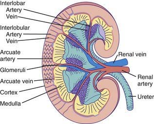
Figure 502-1 Gross morphology of the renal circulation.
(From Pitts RF: Physiology of the kidney and body fluids, ed 3, Chicago, 1974, Year Book Medical Publishers.)

Figure 502-2 Comparison of the blood supplies of cortical and juxtamedullary nephrons.
(From Pitts RF: Physiology of the kidney and body fluids, ed 3, Chicago, 1974, Year Book Medical Publishers.)
The blood supply to each kidney usually consists of a main renal artery that arises from the aorta; multiple renal arteries can occur. The main artery divides into segmental branches within the medulla, becoming the interlobar arteries that pass through the medulla to the corticomedullary junction. At this point, the interlobar arteries branch to form the arcuate arteries, which run parallel to the surface of the kidney. Interlobular arteries originate from the arcuate arteries and give rise to the afferent arterioles of the glomeruli. Specialized muscle cells in the wall of the afferent arteriole and specialized distal tubular cells adjacent to the glomerulus (macula densa) form the juxtaglomerular apparatus that controls the secretion of renin. The afferent arteriole divides into the glomerular capillary network, which then recombines into the efferent arteriole (Fig. 502-2). The juxtamedullary efferent arterioles are larger than those in the outer cortex and provide the blood supply, as the vasa recta, to the tubules and medulla.
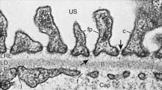
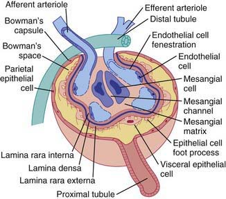
The glomerular network of specialized capillaries serves as the filtering mechanism of the kidney. The glomerular capillaries are lined by endothelial cells (Fig. 502-3) and have very thin cytoplasm that contains many holes (fenestrations). The glomerular basement membrane (GBM) forms a continuous layer between the endothelial and mesangial cells on one side and the epithelial cells on the other. The membrane has 3 layers: (1) a central electron-dense lamina densa; (2) the lamina rara interna, which lies between the lamina densa and the endothelial cells; and (3) the lamina rara externa, which lies between the lamina densa and the epithelial cells. The visceral epithelial cells cover the capillary and project cytoplasmic foot processes, which attach to the lamina rara externa. Between the foot processes are spaces or filtration slits. The mesangium (mesangial cells and matrix) lies between the glomerular capillaries on the endothelial cell side of the GBM and forms the medial part of the capillary wall. The mesangium may serve as a supporting structure for the glomerular capillaries and probably has a role in the regulation of glomerular blood flow, filtration, and the removal of macromolecules (such as immune complexes) from the glomerulus. Bowman’s capsule, which surrounds the glomerulus, is composed of a basement membrane, which is continuous with the basement membranes of the glomerular capillaries and the proximal tubules, and the parietal epithelial cells, which are contiguous with the visceral epithelium (Fig. 502-4).
Fogo AB. Renal pathology. In: Avner ED, Harmon WE, Niaudet P, et al, editors. Pediatric nephrology. ed 6. Heidelberg, Germany: Springer-Verlag; 2009:565-598.
Hunley TE, Kon V, Ichikawa I. Glomerular circulation and function. In: Avner ED, Harmon WE, Niaudet P, et al, editors. Pediatric nephrology. ed 6. Heidelberg, Germany: Springer-Verlag; 2009:31-64.


