Chapter 627 Injuries to the Eye
About 30% of all blindness in children results from trauma. Children and adolescents account for a disproportionate number of episodes of ocular trauma. Boys ages 11-15 yr are the most vulnerable; their injuries outnumber those in girls by a ratio of about 4 : 1. The majority of injuries are related to sports, toy darts, other projectiles, sticks, stones, fireworks, paint balls, and air-powered BB guns. The last causes particularly devastating ocular and orbital injuries. Much of the trauma is avoidable (Chapter 5.1). Any part of the orbit or globe may be affected (Fig. 627-1).
Ecchymosis and Swelling of the Eyelids
Ecchymosis and edema of the eyelids is common after blunt trauma. These are self-limiting, absorb spontaneously, and can be treated with iced compresses and analgesics. Periorbital ecchymosis should prompt careful examination of the eye and surrounding structures for more-serious injury such as orbital bone fracture, intraocular hemorrhage, or rupture of the globe.
Lacerations of the Eyelids
Eyelid lacerations can vary from simple to complex. When evaluating an eyelid laceration, key findings include depth of the laceration, its location, and whether there is involvement of the canaliculus. Most superficial eyelid lacerations may be closed by the primary caregiver, but if it is deep, involves the lid margin, or involves the canaliculus the laceration should be evaluated by an ophthalmologist. The levator muscle is responsible for elevation of the eyelid and runs deep to the skin and orbicularis oculi muscle. If the levator muscle is compromised and this is not recognized at initial repair, ptosis will occur. Therefore, if orbital fat is visible in the laceration, the laceration has compromised the skin, orbicularis oculi, levator, and orbital septum and must be meticulously repaired to prevent ptosis. Eyelid margin involvement also requires careful repair to avoid lid malpostition and notch formation. These can lead to ocular surface problems in the future, resulting in corneal scarring and loss of vision. Lacerations involving the canaliculus require intubation of the nasolacrimal system in addition to repair of the laceration to prevent future tearing problems. Proper primary repair of eyelid lacerations often achieves an outcome superior to secondary repair at a later date. As with any eyelid injury, careful examination of the eye and surrounding tissue is required.
Superficial Abrasions of the Cornea
When the corneal epithelium is scratched, abraded, or denuded, it exposes the underlying epithelial basement layer and superficial corneal nerves. This is accompanied by pain, tearing, photophobia, and decreased vision. Corneal abrasions are detected by instilling fluorescein dye and inspecting the cornea using a blue-filtered light. A slit lamp is ideal for this examination, but a direct ophthalmoscope with blue filter or a hand-held Wood lamp is adequate for young children.
Treatment of a corneal abrasion is directed at promoting healing and relieving pain. Abrasions are treated with frequent applications of a topical antibiotic ointment until the epithelium is completely healed. The use of a semipressure patch does not improve healing time or decrease pain. An improperly applied patch can itself abrade the cornea. A topical cycloplegic agent (cyclopentolate hydrochloride 1%) can relieve the pain from ciliary spasm in patients with large abrasions. Topical anesthetics should not be given at home because they retard epithelial healing and inhibit the natural blinking reflex.
Foreign Body involving the Ocular Surface
A foreign body usually produces acute discomfort, lacrimation, and inflammation. Most foreign bodies can be detected by examination in good light with the aid of magnification or a direct ophthalmoscope set on a high plus lens (+10 or +12). In many cases, slit-lamp examination is necessary, especially if the particle is deep or metallic. Some conjunctival foreign bodies tend to lodge under the upper eyelid, causing the sensation of corneal foreign body as they come into contact with the globe on eyelid movement; they can also produce vertically oriented linear corneal abrasions (Fig. 627-2). Finding these abrasions should lead to a suspicion of such a foreign body, and eversion of the lid may be necessary (Chapter 611). If a foreign body is suspected but not found, further examination is indicated. If the history suggests injury with a high-velocity particle, radiologic examination of the eye may be needed to explore the possibility of an intraocular foreign body.

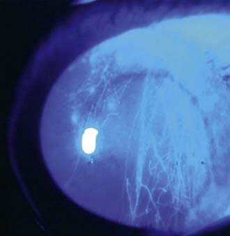
Figure 627-2 Vertically oriented linear corneal abrasions secondary to a foreign body underneath the upper eyelid.
Removal of a foreign body can be facilitated by instillation of a drop of topical anesthetic. Many foreign bodies can be removed by irrigating or by gently wiping them away with a moistened cotton-tipped applicator. Embedded foreign bodies or foreign bodies in the central cornea should be treated by an ophthalmologist. Removal of corneal foreign bodies can leave epithelial defects, which are treated as corneal abrasions. Metallic foreign bodies can cause rust to form in the corneal tissues; examination by an ophthalmologist 1 or 2 days after removal of a foreign body is recommended because a rust ring might require further treatment (curettage).
Hyphema
Hyphema is the presence of blood in the anterior chamber of the eye. It can occur with either a blunt or perforating injury and represents a potential vision-threatening situation. Hyphema appears as a bright or dark red fluid level between the cornea and iris or as a diffuse murkiness of the aqueous humor. Children with hyphema present with acute loss of vision and pain.

Stay updated, free articles. Join our Telegram channel

Full access? Get Clinical Tree

