Inflammatory Bowel Disease

Chapter 328 Inflammatory Bowel Disease


The term inflammatory bowel disease (IBD) is used to represent 2 distinctive disorders of idiopathic chronic intestinal inflammation: Crohn disease and ulcerative colitis. Their respective etiologies are poorly understood, and both disorders are characterized by unpredictable exacerbations and remissions. The most common time of onset of IBD is during the preadolescent/adolescent era and young adulthood. A bimodal distribution has been shown with an early onset at 10-20 yr of age and a 2nd, smaller peak at 50-80 yr of age. About 25% of patients present before 20 yr of age. IBD may begin as early as the 1st yr of life, and an increased incidence among young children has been observed since the turn of the century. Children with early-onset IBD are more likely to have colonic involvement. In developed countries, these disorders are the major causes of chronic intestinal inflammation in children beyond the 1st few yr of life. A 3rd, less-common category, indeterminate colitis, represents ∼10% of pediatric patients.


Genetic and environmental influences are involved in the pathogenesis of IBD. The prevalence of Crohn disease in the United States is much lower for Hispanics and Asians than for whites and blacks. The risk of IBD in family members of an affected person has been reported in the range of 7-30%; a child whose parents both have IBD has a >35% chance of acquiring the disorder. Relatives of a patient with ulcerative colitis have a greater risk of acquiring ulcerative colitis than Crohn disease, whereas relatives of a patient with Crohn disease have a greater risk of acquiring this disorder; the 2 diseases can occur in the same family. The risk of occurrence of IBD among relatives of patients with Crohn disease is somewhat greater than for patients with ulcerative colitis.


The importance of genetic factors in the development of IBD is noted by a higher chance that both twins will be affected if they are monozygotic rather than dizygotic. The concordance rate in twins is higher in Crohn disease (36%) than in ulcerative colitis (16%). Genetic disorders that have been associated with IBD include Turner syndrome, the Hermansky-Pudlak syndrome, glycogen storage disease type Ib, and various immunodeficiency disorders. In 2001 the first IBD gene, NOD2, was identified through association mapping. A few months later, the IBD 5 risk haplotype was identified. These early successes were followed by a long period without notable risk factor discovery. Since 2006, the year of the first published genome wide array study on IBD, there has been an exponential growth in the set of validated genetic risk factors for IBD.


A perinuclear antineutrophil antibody (pANCA) is found in ∼70% of patients with ulcerative colitis compared with <20% of those with Crohn disease and is believed to represent a marker of genetically controlled immunoregulatory disturbance. About 55% of those with Crohn disease are positive for anti-Saccharomyces cerevisiae (ASCA) antibody. Additional markers including antibody to Escherichia coli outer membrane porin (anti-OmpC) and anti-flagellin (anti-CBir1) antibodies are associated with Crohn disease.


Environmental factors are also important and presumably explain discordance between twins and changes in risk among the same race in different geographic regions; the precise factors remain unknown. Persons migrating to developed countries often appear to acquire the higher rates of IBD associated with these regions. Cigarette smoking is a risk factor for Crohn disease but paradoxically protects against ulcerative colitis. No specific infectious agent has been reproducibly associated with IBD, although this is an active area of investigation.


An abnormality in intestinal mucosal immunoregulation may be of primary importance in the pathogenesis of IBD. The gut is under constant immunologic stimulation from microbial agents and dietary antigens. In response, the mucosa normally displays “physiologic” inflammation. In IBD, the mechanisms that keep “physiologic” inflammation in check fail and pathologic inflammation ensues. It is not clear if this represents an abnormal response to customary enteric antigens or a normal response to an as-yet-unidentified microbe. Mediators of inflammation (cytokines, arachidonic acid metabolites, reactive oxygen metabolites, growth factors) are involved, leading to tissue destruction and remodeling with fibrosis. Most therapies are aimed at interfering with these mediators.


It is usually possible to distinguish between ulcerative colitis and Crohn disease by the clinical presentation and radiologic, endoscopic, and histopathologic findings (Table 328-1). It is not possible to make a definitive diagnosis in ∼10% of patients with chronic colitis; this disorder is called indeterminate colitis. Occasionally, a child initially believed to have ulcerative colitis on the basis of clinical findings is subsequently found to have Crohn colitis. This is particularly true for the youngest patients, because Crohn disease in this patient population can more often manifest as exclusively colonic inflammation, mimicking ulcerative colitis. The medical treatments of Crohn disease and ulcerative colitis overlap.


Table 328-1 COMPARISON OF CROHN DISEASE AND ULCERATIVE COLITIS


FEATURE CROHN DISEASE ULCERATIVE COLITIS
Rectal bleeding Sometimes Common
Diarrhea, mucus, pus Variable Common
Abdominal pain Common Variable
Abdominal mass Common Not present
Growth failure Common Variable
Perianal disease Common Rare
Rectal involvement Occasional Universal
Pyoderma gangrenosum Rare Present
Erythema nodosum Common Less common
Mouth ulceration Common Rare
Thrombosis Less common Present
Colonic disease 50-75% 100%
Ileal disease Common None except backwash ileitis
Stomach-esophageal disease More common Chronic gastritis can be seen
Strictures Common Rare
Fissures Common Rare
Fistulas Common Rare
Toxic megacolon None Present
Sclerosing cholangitis Less common Present
Risk for cancer Increased Greatly increased
Discontinuous (skip) lesions Common Not present
Transmural involvement Common Unusual
Crypt abscesses Less common Common
Granulomas Common None
Linear ulcerations Uncommon Common

Extraintestinal manifestations occur slightly more commonly with Crohn disease than with ulcerative colitis (Table 328-2). Growth retardation is seen in 15-40% of children with Crohn disease at diagnosis. Of the extraintestinal manifestations that occur with IBD, joint, skin, eye, mouth, and hepatobiliary involvement tend to be associated with colitis, whether ulcerative or Crohn. The presence of some manifestations, such as peripheral arthritis, erythema nodosum, and anemia, correlates with activity of the bowel disease. Activity of pyoderma gangrenosum correlates less well with activity of the bowel disease, whereas sclerosing cholangitis, ankylosing spondylitis, and sacroiliitis do not correlate with intestinal disease. Arthritis occurs in 3 patterns: migratory peripheral arthritis involving primarily large joints, ankylosing spondylitis, and sacroiliitis. The peripheral arthritis of IBD tends to be nondestructive. Ankylosing spondylitis begins in the 3rd decade and occurs most commonly in patients with ulcerative colitis who have the human leukocyte antigen B27 phenotype. Symptoms include low back pain and morning stiffness; back, hips, shoulders, and sacroiliac joints are typically affected. Isolated sacroiliitis is usually asymptomatic but is common when a careful search is performed. Among the skin manifestations, erythema nodosum is most common. Patients with erythema nodosum or pyoderma gangrenosum have a high likelihood of having arthritis as well. Glomerulonephritis, uveitis, and a hypercoagulable state are other rare manifestations that occur in childhood. Cerebral thromboembolic disease has been described in children with IBD.


Table 328-2 EXTRAINTESTINAL COMPLICATIONS OF INFLAMMATORY BOWEL DISEASE


MUSCULOSKELETAL


SKIN AND MUCOUS MEMBRANES


DERMATOLOGIC


OCULAR


BRONCHOPULMONARY


CARDIAC


MALNUTRITION


HEMATOLOGIC


RENAL AND GENITOURINARY


PANCREATITIS


HEPATOBILIARY


ENDOCRINE AND METABOLIC


NEUROLOGIC


G6PD, glucose-6-phosphate dehydrogenase; IBD, inflammatory bowel disease; PSC, primary sclerosing cholangitis.


Modified from Kugathasan S: Diarrhea. In Kliegman RM, Greenbaum LA, Lye PS, editors: Practical strategies in pediatric diagnosis and therapy, ed 2, Philadelphia, 2004, Saunders, p 285.


328.1 Chronic Ulcerative Colitis


Ulcerative colitis, an idiopathic chronic inflammatory disorder, is localized to the colon and spares the upper gastrointestinal (GI) tract. Disease usually begins in the rectum and extends proximally for a variable distance. When it is localized to the rectum, the disease is ulcerative proctitis, whereas disease involving the entire colon is pancolitis. Approximately 50-80% of pediatric patients have extensive colitis, and adults more commonly have distal disease. Ulcerative proctitis is less likely to be associated with systemic manifestations, although it may be less responsive to treatment than more-diffuse disease. About 30% of children who present with ulcerative proctitis experience proximal spread of the disease. Ulcerative colitis has rarely been noted to present in infancy. Dietary protein intolerance can easily be misdiagnosed as ulcerative colitis in this age group. Dietary protein intolerance (cow’s milk protein) is a transient disorder; symptoms are directly associated with the intake of the offending antigen.


The incidence of ulcerative colitis has remained relatively constant, in contrast to an increase in Crohn disease, but varies with country of origin. Incidence rates are highest in northern European countries and the United States (15/100,000) and lowest in Japan and South Africa (1/100,000). The incidence of ulcerative colitis in Israel varies with the country of origin; those born in Asia or Africa have the lowest risk. The prevalence of ulcerative colitis in northern European countries and the United States varies from 100 to 200/100,000 population. Men are slightly more likely to acquire ulcerative colitis than are women; the reverse is true for Crohn disease.


Clinical Manifestations


Blood, mucus, and pus in the stool as well as diarrhea are the typical presentation of ulcerative colitis. Constipation may be observed in those with proctitis. Symptoms such as tenesmus, urgency, cramping abdominal pain (especially with bowel movements), and nocturnal bowel movements are common. The mode of onset ranges from insidious with gradual progression of symptoms to acute and fulminant. Fever, severe anemia, hypoalbuminemia, leukocytosis, and more than 5 bloody stools per day for 5 days define fulminant colitis. Chronicity is an important part of the diagnosis; it is difficult to know if a patient has a subacute, transient infectious colitis or ulcerative colitis when a child has had 1-2 weeks of symptoms. Symptoms beyond this duration often prove to be secondary to IBD. Anorexia, weight loss, and growth failure may be present, although these complications are more typical of Crohn disease.


Extraintestinal manifestations that tend to occur more commonly with ulcerative colitis than with Crohn disease include pyoderma gangrenosum, sclerosing cholangitis, chronic active hepatitis, and ankylosing spondylitis. Iron deficiency can result from chronic blood loss as well as decreased intake. Folate deficiency is unusual but may be accentuated in children treated with sulfasalazine, which interferes with folate absorption. Chronic inflammation and the elaboration of a variety of inflammatory cytokines can interfere with erythropoiesis and result in the anemia of chronic disease. Secondary amenorrhea is common during periods of active disease.


The clinical course of ulcerative colitis is marked by remission and relapse, often without apparent explanation. After treatment of initial symptoms, ∼5% of children with ulcerative colitis have a prolonged remission (>3 yr). About 25% of children presenting with severe ulcerative colitis require colectomy within 5 yr of diagnosis, compared with only 5% of those presenting with mild disease. It is important to consider the possibility of enteric infection with recurrent symptoms; these infections can mimic a flare-up or actually provoke a recurrence. The use of nonsteroidal anti-inflammatory drugs is considered by some to predispose to exacerbation.


It is generally believed that the risk of colon cancer begins to increase after 8-10 yr of disease and can then increase by 0.5-1% per yr. The risk is delayed by ∼10 yr in patients with colitis limited to the descending colon. Proctitis alone is associated with virtually no increase in risk over the general population. Because colon cancer is usually preceded by changes of mucosal dysplasia, it is recommended that patients who have had ulcerative colitis for >10 yr be screened with colonoscopy and biopsies every 1-2 yr. Although this is the current standard of practice, it is not clear if morbidity and mortality are changed by this approach. Two competing concerns about this plan of management remain unresolved: The original studies may have overestimated the risk of colon cancer and, therefore, the need for surveillance has been overemphasized; and screening for dysplasia might not be adequate for preventing colon cancer in ulcerative colitis if some cancers are not preceded by dysplasia.


Differential Diagnosis


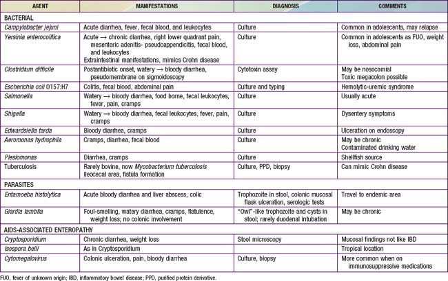
The major conditions to exclude are infectious colitis, allergic colitis, and Crohn colitis. Every child with a new diagnosis of ulcerative colitis should have stool cultured for enteric pathogens, stool evaluation for ova and parasites, and perhaps serologic studies for amebae (Table 328-3). In the setting of antibiotic use, pseudomembranous colitis secondary to Clostridium difficile should be considered. Cytomegalovirus infection can mimic ulcerative colitis or be associated with an exacerbation of existing disease. The most difficult distinction is from Crohn disease because the colitis of Crohn disease can initially appear identical to that of ulcerative colitis, particularly in younger children. The gross appearance of the colitis or development of small bowel disease eventually leads to the correct diagnosis; this can occur years after the initial presentation.


At the onset, the colitis of hemolytic-uremic syndrome may be identical to that of early ulcerative colitis. Ultimately, signs of microangiopathic hemolysis (the presence of schistocytes on blood smear), thrombocytopenia, and subsequent renal failure should confirm the diagnosis of hemolytic-uremic syndrome. Although Henoch-Schönlein purpura can manifest as abdominal pain and bloody stools, it is not usually associated with colitis. Behçet disease can be distinguished by its typical features (Chapter 155). Other considerations are radiation proctitis, viral colitis in immunocompromised patients, and ischemic colitis (Table 328-4). In infancy, dietary protein intolerance can be confused with ulcerative colitis, although the former is a transient problem that resolves on removal of the offending protein, and ulcerative colitis is extremely rare in this age group. Hirschsprung disease can produce an enterocolitis before or within months after surgical correction; this is unlikely to be confused with ulcerative colitis.


Jun 18, 2016 | Posted by in PEDIATRICS | Comments Off on Inflammatory Bowel Disease

Full access? Get Clinical Tree

Get Clinical Tree app for offline access