Infertility and Recurrent Pregnancy Loss


Infertility and Recurrent Pregnancy Loss


Alexander M. Quaas


INFERTILITY

Infertility is defined as the inability to conceive after 12 or more months of regular intercourse without contraception.1 Infertility is common—approximately 10% of couples have difficulty conceiving a child— and has important psychological, economic, demographic, and medical implications.2 Major advances in assisted reproductive treatment technologies have occurred over the last few decades. However, an accurate assessment of factors affecting the fertility of both partners is the initial step to provide targeted treatment. The elements of a standard infertility evaluation are evolving as the available evidence base for commonly performed diagnostic procedures and tests is being reviewed.

Over the last few decades, societal factors affected rates of infertility related to aging in women.

In the United States, there have been increases in the mean maternal age at first birth (25.1 years in 2002 versus 21.4 years in 1968) and in the mean age of women delivering a child (27.3 years in 2002 versus 24.9 years in 1968).3 In part, this trend may be explained by an increasing emphasis on career and educational goals. In addition, a later mean age at first marriage has been observed in countries like England and the United States.4 The advent of widely accessible methods of birth control may also be contributing to a delay in childbirth by allowing earlier unplanned pregnancies to be avoided. With the increased availability of services and improved diagnostic and therapeutic management, more couples are now able to access infertility services.


EPIDEMIOLOGY (OF INFERTILITY)

A couple is said to be infertile if they have been trying to achieve a pregnancy for more than 1 year without success.1 The roots for this somewhat arbitrary definition lie in a study from 1956 analyzing a cohort of 5574 English and American women engaging in unprotected intercourse who ultimately conceived over a 10-year period.5 In this study, 50% conceived within 3 months, 72% within 6 months, and 85% within 12 months.

The definition can also be justified from a statistical perspective using the concepts of fecundity and cumulative pregnancy rates.

Fecundity (f) represents the probability of conception per month of effort and is calculated by dividing the number of conceptions (C) by the person-months of exposure (T).

f = C/T

It must be remembered that even in infertile populations, fecundity is almost never zero, and for fertile couples, it is approximately 0.20. Assuming a 20% monthly probability of pregnancy among normally fertile couples, the cumulative probability of pregnancy after 12 months is 93%.6 Therefore, there is a 7% statistical probability that a normally fertile couple does not conceive after 12 months, close to the 5% statistical threshold for a type I error (in this case, a false rejection of the null hypothesis of normal fertility). The 1-year definition of infertility is thus both statistically and clinically meaningful. Therefore, it is reasonable to perform a full infertility evaluation only when a couple has attempted 12 months of unprotected intercourse. With increasing age, however, it is common practice to not delay the workup given the rapid decrease in ovarian reserve. Therefore, many specialists recommend testing for common causes of infertility after 6 months in a couple with a female older than age 35 years and immediately in a couple with female older than age 40 years.7

Infertility can be considered as a reduction in fecundity to less than that of the general population, and the evaluation is designed to identify factors that are responsible for diminished fecundity. Based on a woman’s history, infertility is classified into two categories: primary and secondary. Primary infertility implies no antecedent pregnancy, and secondary infertility is defined by a history of any pregnancy, including abortions and ectopic pregnancies.

Estimating the prevalence of infertility is difficult because of inconsistent definitions in epidemiologic reports. Between 1965 and 1988, the prevalence appeared to remain stable at approximately 13% of American women aged 15 to 44 years (excluding surgically sterilized individuals).8,9 Although the prevalence of infertility has remained at approximately 13 to 15% of the U.S.
population since the mid-1980s, more couples are seeking medical and surgical services for impaired fecundity. The number of childless women who are 35 to 44 years old increased by more than 1 million from 1982 to 1988. Women were 2 to 3 years older in 1990 than in 1979 when they delivered their first child, and significantly, more women never had a child.10

Differences in the distribution of primary and secondary infertility have a direct impact on the use of fertility services because women with primary infertility are more likely to use medical services than women with secondary infertility. Overall, about half of all infertile couples will seek treatment, although only 25% will obtain assistance from an infertility specialist.11 Geographic variations in the use of infertility services are in part mediated by differences in state-mandated insurance coverage of assisted reproductive services.12 Approximately half of couples with infertility eventually conceive. Likelihood of conception is influenced by several factors, including the duration of infertility, the causes of infertility, and the age of the woman at the time of treatment.

In a World Health Organization (WHO) study of 8500 infertile couples, the distribution of medical conditions contributing to infertility was analyzed.13 In developed countries, female factor infertility was reported in 37% of infertile couples, male factor infertility in 8%, and both male and female factor infertility in 35%. Five percent of couples had unexplained infertility and 15% became pregnant during the study. This study illustrates the importance of evaluating both partners of the infertile couple, focusing on the major processes which affect fertility.


INVESTIGATION OF THE INFERTILE COUPLE

Clinical evaluation of the infertile couple is customarily initiated if a pregnancy has not occurred after 1 year of regular unprotected intercourse. Earlier assessment is warranted in women who have a history of oligomenorrhea/amenorrhea, are older than age 35 years, and have known or suspected pelvic pathology.14 Initially, a thorough history and physical exam are essential to guide the initial individual diagnostic approach. The history should include details on patient age, the duration of infertility, results of any previous evaluations, and a menstrual history to help determine ovulatory status.15 A sexual history, including sexual dysfunction and frequency of coitus, should be obtained, as well as a complete medical, surgical, gynecologic, and obstetric history. A personal and lifestyle history is essential, given that factors such as occupation, exercise, stress, dieting, smoking, as well as drug and alcohol use, can affect fertility. For example, smoking or other tobacco use, marijuana, and cocaine may reduce fecundity in men and women.16 Occupational and environmental exposures associated with decreased fecundity are reviewed in a separate chapter. Some centers find it helpful to distribute a detailed questionnaire to patients before their first visit to obtain information that is sometimes difficult to express in an office interview. Such a questionnaire is available from the American College of Obstetricians and Gynecologists (ACOG) or the American Society for Reproductive Medicine (ASRM).

The physical exam may identify potential causes of infertility and previously undiagnosed health conditions. For example, a goiter may be palpated in a patient with underlying thyroid disease, and male pattern baldness may be found in a female patient with androgen excess. The pelvic exam may reveal tenderness or masses in the adnexa or posterior cul de sac, suggesting endometriosis.

After the initial evaluation, further diagnostic assessment can be planned. As part of preconception evaluation and counseling, a rubella titer should be ordered, indicated immunizations should be administered, HIV testing, and cystic fibrosis (CF) carrier testing should be offered to all couples. Prenatal vitamins containing adequate amounts of folic acid should also be prescribed at this time to decrease the risk of fetal neural tube defects.17 Serum thyroid-stimulating hormone (TSH) testing is performed because undiagnosed maternal hypothyroidism is associated not only with anovulatory infertility but also with adverse fetal neuropsychological development.18

A key objective of further testing is to rule out male factor infertility, anovulation, or tubal obstruction7 (see Table 11.1). Most couples will require semen analysis, an assessment of ovulatory function, an assessment of the uterine cavity, and an assessment of tubal patency by hysterosalpingography or laparoscopy. Ovarian reserve testing by estimation of follicle-stimulating hormone (FSH) and estradiol on cycle day 3 is often ordered as part of the initial testing. Laparoscopy may be considered in a minority of cases when endometriosis, intrapelvic adhesions, or fallopian tube disease is suspected.

Expert committees may offer guidance in the initial evaluation of the infertile couple such as the ASRM Practice Committee (www.asrm.org).19


TABLE 11.1 Assessment of Infertility


Initial tests for couples with infertility:


  • Semen analysis (to assess any male factor component)


  • Menstrual history, assessment of LH surge in urine prior to ovulation, and/or luteal phase progesterone level (to evaluate ovulatory function)


  • Hysterosalpingography (to determine tubal patency and the status of the uterine cavity)


  • Day 3 serum FSH and estradiol levels (to evaluate ovarian reserve)


In some instances, the following additional tests may be indicated:


  • Pelvic ultrasound (to look for uterine myomas and ovarian cysts)


  • Laparoscopy (to determine if there is endometriosis or other pelvic pathology)


  • Further assessment of ovarian reserve in women older than age 35 years; this may involve a clomiphene citrate challenge test, ultrasound for early follicular antral follicle count, day 3 serum inhibin B level, or anti-Müllerian hormone measurement


  • Thyroid function testing


LH, luteinizing hormone; FSH, follicle-stimulating hormone.


ANOVULATORY INFERTILITY

Ovulatory dysfunction and anovulation affect 15 to 25% of all infertile couples seeking therapy. The ultimate proof of normal ovulation is a subsequent pregnancy, but several other pieces of evidence can be used to confirm the presence or absence of normal ovulation. The process leading to ovulation is composed of several components; disruption of any of these can impede ovulation or the capacity of a mature oocyte to fertilize. Treatment of ovulation disorders remains one of the most successful of all infertility treatments.


Evaluation of Ovulatory Function


History

The first and arguably most telling piece of information about ovulatory function is a thorough menstrual history. Generally speaking, regular menstrual cycles between 28 and 35 days are ovulatory. Shorter or longer cycles may signify anovulation; in shorter cycles, this may be representative of a shortened follicular phase seen in aging, and longer cycles may belie an inadequate luteal phase resulting in an endometrium unreceptive to an embryo. A patient who reliably reports very regular 28-day menstrual cycles is unlikely to have anovulation. On the other hand, a woman who reports infrequent, irregular menses and no moliminal symptoms (or who gives an unclear history of her menstrual cycles) is likely oligo- or anovulatory and should be evaluated.


Background: Physiology of Normal Ovulation

A review of the normal hormonal events that regulate the ovulatory process is essential to an understanding of the tests devised for ovulation detection. The trophic effects of both FSH and luteinizing hormone (LH) result in maturation of ovarian follicles. Release of both FSH and LH from the anterior pituitary is controlled by gonadotropin-releasing hormone (GnRH), which is produced in the hypothalamus in a pulsatile fashion. The ovulatory process is characterized by a rapid midcycle rise in LH that culminates in the LH peak. A consensus from previous reports places the onset of the serum LH surge approximately 36 to 38 hours before ovulation.20 FSH also increases midcycle but to a lesser degree than LH. The luteal phase is characterized by a rise in the concentration of progesterone produced by the corpus luteum, with maximal concentration reached about 8 days after the LH peak. The menstrual cycle length is variable, with the variation residing in the follicular phase. The length of the luteal phase is consistently 14 days.


Midluteal Serum Progesterone Testing

Detection of serum progesterone greater than 3 ng/mL on day 21 suggests the presence of a functioning corpus luteum, indicating that ovulation occurred.21 One report indicated that midluteal progesterone levels greater than 8.8 ng/mL were present in more than 95% of spontaneous conception cycles.22 Given the large variability in progesterone secretion throughout the luteal phase in normal patients, it is controversial whether a higher level of progesterone indicates a “better” ovulation than a lower level.


Urinary Luteinizing Hormone Testing

Detection of an LH surge, suggesting that ovulation is imminent, allows the patient to appropriately time intercourse.23 There is a 4- to 6-hour lag between serum and urinary LH surges. More than 90% of ovulation episodes can be detected by a single urinary test performed midafternoon or early evening of the day of the surge. This helps couples to define the interval in which conception is most likely (the 2 days prior to ovulation and the third day or day of ovulation).
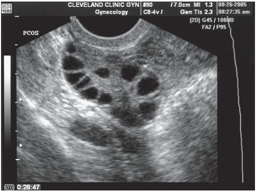
24 Accuracy, reliability, and ease of use vary among the different LH detector kits. These kits are helpful for women in whom ovulation is known to occur; false positives may result in women with polycystic ovarian syndrome (PCOS), premature ovarian failure, and menopause—all conditions associated with increased LH levels.


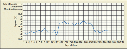
Basal Body Temperature Charting

Basal body temperature (BBT) charting has long been used because of its simplicity. The patient is asked to record her body temperature first thing in the morning from the same site each day throughout her menstrual cycle and chart it on a BBT chart (Fig. 11.1).

The increases in progesterone associated with ovulation will lead to an increase in BBT in the days following ovulation. Biphasic patterns are characteristic in ovulatory cycles; monophasic recordings or a grossly short interval of luteal phase temperature elevation (less than 11 days) may identify patients with absent or poor quality ovulatory function. Challenges to the use of this technique are its need for a special basal body thermometer where differences of one-tenth of a degree may be easily discerned, its inconvenience (the woman must take her temperature first thing upon awakening and doing anything such as urinating, eating or drinking, and variability—some ovulatory women may exhibit monophasic BBT patterns), and the test cannot reliably define the time of ovulation.25 The temperature rise seen with ovulation starts 1 to 2 days after the LH surge and lasts for 10 or more days, but because ovulation has already occurred once the increase is seen, it is too late to use the BBT chart for timing sexual intercourse for conception.


Endometrial Biopsy

Histologic evidence of secretory endometrium detected by endometrial biopsy is indicative of ovulation and corpus luteum formation. The endometrial biopsy is performed in the midluteal phase, around 7 days prior
to the expected period. Although the test has been used to diagnose “luteal phase deficiency” in the past, experts have moved away from this concept because of a lack of intra- and interobserver consistency; and the test is unable to provide further useful information other than the presence of ovulation.26 Further disadvantages of endometrial sampling include expense, patient discomfort, and the known risks of the procedure.


FIGURE 11.1 Example of a basal body temperature (BBT) recording chart. (From Hyde J, DeLamater J. Understanding Human Sexuality. 6th ed. New York: McGraw-Hill; 1997.)


Transvaginal Ultrasound Monitoring

The use of serial transvaginal sonography is a reliable technique to monitor follicular development. Ovulation is deemed to have occurred if the follicle reaches a mean diameter of 18 to 25 mm and subsequently changes in sonographic density or demonstrates sonographic evidence of follicular collapse. However, its use as an initial diagnostic test for all infertile women is not cost-effective.


Other Evaluation: Special Tests to Determine the Underlying Etiology

Once oligo- or anovulation has been diagnosed, additional tests aim to identify the etiology and help choose the optimal treatment. The most common causes of ovulatory dysfunction are PCOS, thyroid dysfunction, hyperprolactinemia, and late-onset congenital adrenal hyperplasia. Therefore, evaluation for PCOS should be undertaken as well as determinations of serum TSH, prolactin, and 17-hydroxyprogesterone.

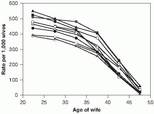
Prior to instituting treatment for oligo- or anovulation, the patient undergoes ovarian reserve testing, which includes the measurement of an early follicular phase serum FSH and estradiol level. As women age, changes in the patterns and levels of gonadotropin release occur. The follicular phase shortens, and this is associated with an increase in early follicular phase serum FSH prior to any noticeable changes in peak estradiol or progesterone levels or changes in the luteal phase. A high level of FSH on day 3 (e.g., greater than 10 mIU/mL) of the cycle is associated with a poor response to in vitro fertilization (IVF), although a single measurement may not be sufficient—usually two or more high levels in different cycles is required to truly be reliable as a prognostic indicator for IVF response. It is important to note that different FSH assays may yield large variations in measurements even in the same blood sample, so it is important that providers be aware of which assay is being used if specimens go to different labs or try to send all specimens to the same lab for analysis with the same assay.

A high serum estradiol (e.g., greater than 80 pg/mL) is also associated with a poor prognosis for pregnancy after IVF and is usually associated with accelerated, premature follicle recruitment and a reduction in available oocytes. Like FSH measurements, estradiol levels vary widely with different assays.

Other tests for ovarian reserve include anti-Müllerian hormone (AMH) levels, inhibin B levels, clomiphene citrate challenge test (CCCT), exogenous FSH ovarian reserve test, and the use of ultrasound to determine antral follicle count or ovarian volume.


Treatment of Anovulatory Infertility

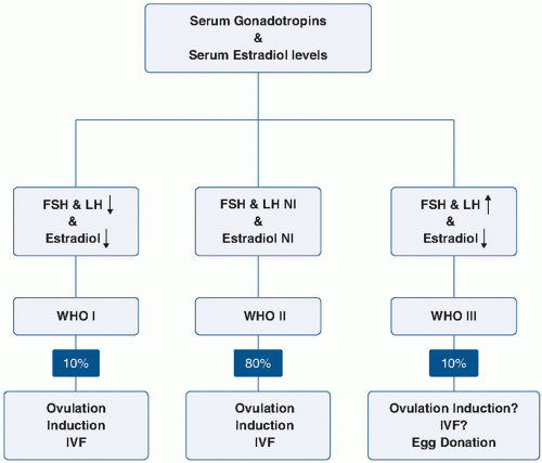
WHO has classified anovulation into three main groups (Fig. 11.2; Table 11.2). Women in WHO Group I have hypogonadotropic hypogonadism or hypothalamic amenorrhea. Common causes of this include central nervous system disorders such as hypothalamic or pituitary diseases, stress, eating disorders, and exercise-induced ovulatory dysfunction. Eighty percent of patients with ovulatory dysfunction will have estradiol and gonadotropin levels in the normal range and fall into the category of WHO II, eugonadotropic ovulatory dysfunction.27
(Group II is discussed in more detail later.) Group III includes hypergonadotropic hypogonadism which may be due to primary ovarian insufficiency, commonly referred to as premature ovarian failure, or to gonadal dysgenesis. The WHO also recognizes that hyperprolactinemia anovulation is a separate category, and usually the gonadotropins are normal or decreased in these cases.


FIGURE 11.2 WHO classification of anovulation. (From Laven JS, Joop SE, Fauser BC. What role of estrogens in ovarian stimulation. Maturitas. 2006;54: 356-362.)


TABLE 11.2 World Health Organization Classification of Anovulation


WHO Class 1: Hypogonadotropic Hypogonadal Anovulation (Hypothalamic Amenorrhea)


These women have low or low-normal serum follicle-stimulating hormone (FSH) concentrations and low serum estradiol concentrations due to decreased hypothalamic secretion of gonadotropin-releasing hormone (GnRH) or pituitary unresponsiveness to GnRH. Other causes may include stress, eating disorders, or exercise-induced amenorrhea. Workup may include MRI of hypothalamus and pituitary region of brain.


WHO Class 2: Normogonadotropic Normoestrogenic Anovulation


These women may secrete normal amounts of gonadotropins and estrogens. However, FSH secretion during the follicular phase of the cycle is subnormal. This group includes women with polycystic ovarian syndrome (PCOS). These patients should also be screened for other causes of increased male hormones and also for metabolic disorders associated with the condition such as lipid profile abnormalities and diabetes. Some ovulate occasionally, especially those with oligomenorrhea.


WHO Class 3: Hypergonadotropic Hypoestrogenic Anovulation


The primary causes are premature ovarian failure (absence of ovarian follicles due to early menopause) and ovarian resistance (follicular form). This group may also include hyperprolactinemic anovulation.


Adapted from Agents stimulating gonadal function in the human: report of a WHO Scientific Group. World Health Organ Tech Rep Ser. 1973;514:1-30. http://whqlibdoc.who.int/trs/WHO_TRS_514.pdf. Accessed December 2, 2013.


Group I patients often have low body mass index (BMI) (less than 17 kg/m2) resulting from eating disorders or excessive exercise. Mental and emotional stress may disrupt the pulsatile release of GnRH, which leads to a reduction in pituitary secretion of gonadotropin(s) and impairs ovarian function. Treatment usually involves a modification of diet, weight gain, and/or a reduction in exercise, although these approaches are not always well accepted or adhered to by patients. In patients who fail to respond to lifestyle changes, pulsatile GnRH therapy may be an option, and although it is approved for use in the United States, it is not currently available.

The majority of patients in the WHO II category have PCOS.14 PCOS is characterized by chronic anovulation, hyperandrogenism, and insulin resistance. Diagnostic guidelines vary according to the Rotterdam criteria, the National Institutes of Health, and the Androgen Excess Society, but all include evidence of oligo- or amenorrhea and clinical or biochemical evidence of hyperandrogenism.28

The first line of intervention should be lifestyle change when appropriate because interventions that reduce circulating insulin levels in women with PCOS may restore normal reproductive endocrine function. In patients with BMI greater than 30 kg/m2, weight loss and exercise is the initial recommended treatment for this etiology of anovulation.

If lifestyle intervention alone is ineffective, the first-line agent for ovulation induction in PCOS is clomiphene citrate (CC), an estrogen antagonist that increases gonadotropin release. Clomiphene is usually well tolerated; side effects include hot flushes, mood swings and (rarely) visual changes, and ovarian hyperstimulation syndrome.29 Available evidence suggests that approximately half of the patients will ovulate on an initial CC regimen of 50 mg for 5 days starting on cycle day 3, 4, or 5 (cycle day 1 is the first day of menstruation). If the patient does not respond, the dose may be increased to 100 mg or even 150 mg.14 In patients with very irregular menses, micronized progesterone (200 mg daily for 10 days) or medroxyprogesterone acetate (10 mg daily for 10 days) can be given and clomiphene started on cycle day 3 after initiation of the withdrawal bleed. Documentation of ovulation is made with urinary LH monitoring and/or a luteal progesterone level. If clomiphene is taken on cycle days 3 through 7, ovulation usually occurs between days 12 and 15. Typically, each dosage is tried during one menstrual cycle, with an increase in the dose for the next cycle, but recent evidence suggests that patients may be treated with a “stair-step protocol,” during which the dose is increased during the same ovulation induction cycle, if no follicular activity is noted on ultrasound.30

If CC is not successful, the treatment choices include adjunctive use of insulin-sensitizing agents, aromatase inhibitors, gonadotropin therapy, and laparoscopic ovarian diathermy.

Insulin-sensitizing agents such as metformin (Glucophage) have been shown to increase the frequency of spontaneous ovulation, menstrual cyclicity, and the ovulatory response to clomiphene.31,32 Many women with PCOS who are resistant to CC have demonstrable insulin resistance and hyperinsulinemia. Insulin-sensitizing agents taken alone or in combination with CC can restore ovulation. A recent multicenter trial was designed to study the question whether clomiphene, metformin, or both together should be the first-line therapy in patients with PCOS and infertility.33 Live-birth rates were 22.5% (47 of 209 subjects) in the clomiphene group, 7.2% (15 of 208) in the metformin group, and 26.8% (56 of 209) in the combination-therapy group (p <0.001 for metformin versus both clomiphene and combination therapy; p = 0.31 for clomiphene versus combination therapy). Most experts therefore recommend that clomiphene alone should be the first-line therapy for anovulation in PCOS, with metformin added if there is evidence of hyperinsulinemia or for clomiphene-resistant patients.

Aromatase inhibitors such as letrozole block the conversion of androgens to estrogens and therefore increase gonadotropin concentrations by reducing the negative feedback on the pituitary gland. Letrozole has similar efficacy in ovulation induction as clomiphene, as demonstrated in multiple trials and meta-analyses.34 However, a study from Canada presented as an abstract in 2005 raised concerns over its safety with respect to cardiac malformations, which led to a warning from the manufacturer of the medication. Despite the fact that subsequent studies demonstrated a comparable safety profile of the medication compared to clomiphene,35 practitioners tend to still be cautious about the use of letrozole for ovulation induction.

Treatment with injectable gonadotropins with or without intrauterine insemination (IUI) requires close hormonal and sonographic monitoring, is costly, and has significant potential to result in multiple gestation. Laparoscopic destruction of ovarian follicles by laser or cautery has similar success rates as gonadotropin treatment but with lower multiple pregnancy rates.36 However, risks of the procedure include the formation of adnexal adhesions and a decrease in ovarian reserve. Therefore, this procedure is usually limited to patients for whom other alternatives have been exhausted and patients who do not have access to the most costly but also most effective treatment for clomiphene-resistant PCOS patients, namely IVF.

Premature ovarian failure is defined as the development of primary hypogonadism in a woman prior to the age of 40 years. Some of these women may experience
intermittent ovulation, and 5 to 10% may conceive and deliver.37 Premature ovarian insufficiency may be due to a variety of causes, including chromosomal defects such as Turner syndrome, fragile X permutation carriers, galactosemia, radiation exposure, certain drug exposures, and autoimmune disease. Despite a growing list of mutation associated with premature ovarian insufficiency, the etiology is still unknown in 75 to 90% of cases.38

In about 3% of women, the primary ovarian failure may precede the development of an autoimmune adrenal insufficiency, a potentially fatal disorder, by several years. Proper screening with serum anti-adrenal and anti-21 hydroxylase antibodies is recommended for these patients.39 These patients are also at increased risk for developing autoimmune hypothyroidism and should be screened for this as well. Although ultrasound studies indicate that follicular development occurs frequently in these women, ovulation is infrequent.40 Because spontaneous pregnancy rates are low in this population, treatment options include IVF with oocyte donation and adoption. The evidence to support the use of exogenous estrogen, gonadotropin therapy (with estrogen or with GnRH agonist) are fairly limited and not overly encouraging.41, 42, 43, 44 Success rates for the use of IVF with oocyte donation depend primarily on the age of the oocyte donor.

In patients with elevated prolactin levels, anovulation is secondary to decreased estradiol concentrations. One isolated increased prolactin level should be confirmed by repeat measurement in the early morning, avoiding recent stress or recent breast or pelvic exams, which can cause mild elevations. If the value is confirmed, hypothyroidism should be ruled out, and magnetic resonance imaging is indicated to rule out a pituitary adenoma. Treatment of hyperprolactinemic anovulation, even in the presence of a pituitary adenoma, is usually by means of dopaminergic drugs, such as bromocriptine (Parlodel). Bromocriptine is an ergot alkaloid derivative with dopamine receptor agonist activity that directly inhibits prolactin secretion.

For ovulation induction, bromocriptine is usually started at 1.25 mg orally at bedtime for 1 week and then increased to 2.5 mg twice daily. After 1 week on this dose (2.5 mg), ovulatory status should be evaluated. In the absence of ovulation, the dose can be increased in 1.25-mg increments (the maximum dosage should not exceed 100 mg/day).45 After ovulation is established, the medication is maintained until the patient becomes pregnant. Bromocriptine is usually discontinued after a positive pregnancy test but can be used safely during pregnancy. Most patients who conceive do so within six ovulatory cycles with the average being two cycles.46 Side effects of bromocriptine include nausea, headache, and faintness due to orthostatic hypotension. These effects are minimized by gradually increasing the dose. Taking it with food is recommended to avoid gastrointestinal side effects. Vaginal administration of bromocriptine has been associated with fewer side effects but with similar effectiveness.47

Another dopamine agonist, cabergoline (Dostinex), given once a week, is more effective in normalizing prolactin and restoring menses than bromocriptine and significantly better tolerated. However, given the fact that less data exist on safety in pregnancy, it has not been widely accepted as first-line therapy for ovulation induction in hyperprolactinemic patients seeking fertility.48


DECREASED OVARIAN RESERVE

Female fertility begins to decline many years prior to the onset of menopause despite continued regular ovulatory cycles. This occurs because of diminished oocyte quality, decreased oocyte numbers, and reproductive potential and is referred to as decreased ovarian reserve. In addition to declining fecundity, the risk of spontaneous abortion increases with a woman’s age. Although age-related changes in fecundity have been documented in several populations, there is significant variability in the timing of onset of diminished reproductive potential for individual women. Although IVF was initially discovered as a treatment for tubal factor infertility, a steep rise in the number of cases performed for women older than age 40 years since 1990 made decreased ovarian reserve the most common indication for the use of assisted reproductive technology (ART) in most centers. In order to assess ovarian reserve, multiple factors must be taken into account. The most important factor in this assessment is female age, which has been shown to be inversely associated with fertility in multiple populations49 (Fig. 11.3).


FIGURE 11.3 Marital fertility rates by age group in different populations. (From Committee on Gynecologic Practice of the American College of Obstetricians and Gynecologists; Practice Committee of the American Society for Reproductive Medicine. Age-related fertility decline: a committee opinion. Fertil Steril. 2008;90:S154-S155.)


The laboratory tests that have been used the longest to assess ovarian reserve are day 3 (basal) FSH and estradiol concentrations. Women with normal ovarian reserve are able to produce enough ovarian hormones from the small follicles in the early follicular phase to keep the FSH low. If there are a reduced number of follicles or oocytes, ovarian hormonal production will be low, and this will incur a rise in FSH early in the cycle. Although differences exist between laboratories, follicular phase FSH values greater than 10 mIU/mL or day 3 serum estradiol values greater than 80 pg/mL indicate poor prognosis for conception.50 Because of the variation in FSH values between laboratories depending on the assay and methodology used, cutoff values may range from 10 to 25 mIU/mL. For this reason, clinicians should know the critical values for their respective labs. A single elevated day 3 FSH value implies poor prognosis for pregnancy, even when values in subsequent cycles are normal.51,52

Women with diminished ovarian reserve demonstrate premature recruitment of follicle in the menstrual cycle follicular phase. These elevated estradiol levels may inhibit pituitary FSH production yielding lower FSH levels. Measuring both the FSH and estradiol helps to avoid interpretation of a false-negative FSH test as indicative of adequate ovarian reserve.

The CCCT, another test of ovarian reserve, involves measuring day 3 FSH, administration of clomiphene 100 mg daily from days 5 to 9, and repeat FSH measurement on day 10.53 If either the day 3 or the day 10 level is elevated (greater than 12 IU/L), decreased ovarian reserve can be diagnosed. The rationale behind this test was that women with adequate ovarian reserve should develop a cohort of follicles producing adequate estradiol and inhibin to suppress the FSH. However, given that the test is relatively involved, and with the advent of newer and more sensitive tests of ovarian reserve, the CCCT is rarely used in clinical practice nowadays. If abnormal, the day 3 FSH and CCCT tests are fairly similar in their ability to predict that pregnancy will not occur if the woman’s oocytes are used. If they are normal, they are not particularly useful in predicting fertility, but they do indicate that there is an adequate ovarian reserve.

More and more commonly, ovarian reserve is assessed by measuring serum AMH concentrations.54,55 AMH is a homodimeric disulfide-linked glycoprotein of the transforming growth factor (TGF)-β super-family produced by the granulosa cells of the preantral and small antral follicles.56 Serum AMH concentrations have been found to be a reflection of the size of the primordial follicle pool and are highly correlated with the response to ovarian stimulation as part of ART indicative of ovarian reserve.54,55,57 Serum AMH may be predictive of the age of onset of menopause, and concentrations are thought to be stable throughout the menstrual cycle, although this is under investigation.58,59 It is not detectable at menopause.60 There is no uniform agreement on the appropriate threshold value of AMH diagnostic of diminished ovarian reserve. Levels above 0.5 ng/mL are thought to represent a good reserve, whereas those less than 0.15 ng/mL are indicative of a poor response to IVF if the woman’s oocytes are used.61,62


FIGURE 11.4 Transvaginal ultrasound image demonstrating antral follicles. (Reproduced with permission from Falcone T. Infertility. Disease management project. The Cleveland Clinic Center for Continuing Education. http://www.clevelandclinicmeded.com/medicalpubs/diseasemanagement/womens-health/infertility/. Accessed December 2, 2013. Copyright © 2000-2011 The Cleveland Clinic Foundation. All rights reserved.)

Another less commonly used serum test is the assessment of inhibin B produced by the granulosa cells of ovarian follicles. As the number of developing follicles decreases, there is a parallel decline in the serum level of inhibin B. In early menopause, the early rise in FSH is closely related to the fall in inhibin B and suggests that inhibin B may play a large role in the normal control of FSH release.63

Ultrasound assessment of the antral follicles in both ovaries, known as the antral follicle count (AFC), is another valuable tool when assessing ovarian reserve. Antral follicles have been defined as measuring 2 to 10 mm in the greatest two-dimensional plane64 (Fig. 11.4).

A low AFC (suggested by some to be 4 to 10 antral follicles between days 2 and 4) has been associated with poor response to ovarian stimulation and with the failure to achieve pregnancy.65 Age-related nomograms with normal ranges by age are being developed. Ovarian volume on ultrasound is another finding that has been correlated with ovarian reserve but is less predictive and less commonly used than the AFC.

In clinical practice, there is no one single factor to assess ovarian reserve. An integrated assessment of age as well as laboratory and ultrasound testing will be most useful to guide patient counseling and treatment.


STRUCTURAL AND ANATOMIC CAUSES OF INFERTILITY

Approximately 40% of all infertile women will demonstrate an abnormality of the uterus, cervix, and/or
fallopian tubes. To date, the best assessment of pelvic anatomic causes of infertility is with a thorough history and physical exam as well as a hysterosalpingogram.64 Other investigations are necessary depending on patient characteristics as outlined in the following section.


Cervical Factor Infertility

The long-held belief that abnormalities of cervical mucus production or of sperm-mucus interaction are the sole cause of infertility has recently been questioned.64 If chronic cervicitis is diagnosed on exam and upon examination of the cervical mucus, it should be treated.

Some practitioners still perform postcoital testing (PCT) by assessing the presence of motile sperm in the cervical mucus microscopically shortly after intercourse around the time of ovulation. However, the test has been found to be cumbersome for patients, poorly predictive of outcomes, subjective, and of little impact on clinical decision making. Therefore, it has largely been abandoned in clinical practice.66,67


Uterine Factors

Uterine factors are rarely the sole cause of infertility but may contribute to infertility in many patients. Multiple methods exist to assess the uterine cavity, looking for intracavitary lesions.

The most commonly used method in this context is the use of hysterosalpingogram (HSG). The procedure involves the insertion of 10 to 20 mL of contrast material into the uterus via a balloon catheter. Women who are allergic to shellfish or iodinated contrast agents cannot undergo HSG testing. Serial plain pelvic x-ray examination provides immediate information about the appearance of the endocervical canal, the uterine cavity, and the fallopian tube lumina7 (Fig. 11.5).

Prophylactic antibiotics (doxycycline 100 mg twice a day for 3 days prior to the hysterosalpingogram) are warranted in women with a prior history of pelvic infection, specifically Chlamydia.68 The HSG can reveal Müllerian anomalies such as unicornuate or septate uteri and intracavitary lesions such as endometrial polyps or submucosal fibroids. It can also detect the presence of intrauterine adhesions/synechiae (Asherman syndrome) and was the first imaging modality to diagnose adenomyosis noninvasively.69 However, the sensitivity and the positive predictive value of the HSG to detect the most common lesions, polyps, and fibroids is low.70


FIGURE 11.5 Example of a normal hysterosalpingogram (HSG). (From Baramki TA, Hysterosalpingography. Fertil Steril. 2005;83(6):1595-1606, with permission from Elsevier.)

Saline hysterography involves the insertion of an HSG catheter into the uterine cavity to introduce sterile saline while visualizing with the transvaginal ultrasound71 (Fig. 11.6). This technique is thought to be more sensitive than HSG in the detection of intrauterine pathology, with higher negative and positive predictive values.70

The gold standard for diagnosis and treatment of intracavitary uterine lesions is hysteroscopy, which can be done in the office or in the operating room. Because this technique is more involved and costly, many authors recommend evaluation of the uterine cavity with HSG
and saline hysterography as first-line evaluation of the infertile patient, followed by hysteroscopic evaluation if abnormalities are detected.64,72


FIGURE 11.6 Example of a hydrosonogram used to detect and delineate an intracavitary lesion. (From Williams CD, Marshburn PB. A prospective study of transvaginal hydrosonography in the evaluation of abnormal uterine bleeding. Am J Obstet Gynecol. 1998;179:292-298.)

Regarding therapy of intrauterine lesions, most practitioners aim to restore normal anatomy and a pristine uterine cavity prior to pursuing conception spontaneously or using ART. This concept has been well documented and studied for some conditions, whereas the evidence is weaker for other lesions such as uterine polyps. Currently, there is good evidence that intrauterine adhesions should be resected73 and that submucosal myomas should be removed.74 Intramural fibroids that are not impinging on the cavity but are large (greater than 5 cm in diameter) should also be removed prior to IVF.75 According to the theory that endometrial polyps may act as intrauterine devices and impede implantation, most specialists remove any polyps found on evaluation prior to ART, even though evidence for this concept from large well-designed studies is lacking.


Tuboperitoneal Factors

The most common etiology for tubal disease causing infertility is pelvic inflammatory disease (PID), most often secondary to genital infection with Chlamydia trachomatis. The bacteria cause damage to the ciliated epithelium, resulting in impaired tubal transport. Many cases go undetected and untreated because 50 to 80% of women with these infections are asymptomatic.

Pelvic adhesions secondary to endometriosis or previous abdominal surgery are other etiologies of tubal distortion and dysfunction.

Therefore, historically, attempts were made to select which patients should be referred for a hysterosalpingogram (HSG) based on history, such as history of PID, pelvic surgery, or ruptured appendix, duration of infertility and infertility treatment, as well as patient age. Testing for antichlamydial antibodies was introduced to screen for and detect tubal pathology. It was shown that positive serology was moderately effective at predicting the risk of finding tubal pathology on HSG and laparoscopy, with a high negative predictive value.76, 77, 78 However, in the evolution of the basic infertility evaluation over the years, the Chlamydia antibody test was found to have less and less clinical use because of its modest sensitivity for tubal disease and because of the increasing recommendation that every patient presenting for infertility evaluation should undergo HSG testing.64 Additionally, PID can result from other pathogens, and tuboperitoneal pathology can result from other causes.

Therefore, HSG testing is the mainstay of evaluation for tubal pathology. Spill of contrast into the peritoneal cavity should be documented (see Fig. 11.5). Collection of dye around the distal tube that does not spill into the cavity with changing patient position may suggest paratubal adhesions. When tubal blockage is diagnosed on HSG, the location of the blockage is crucial. Unilateral proximal blockade with a normal contralateral tube is most often due to tubal spasm. Distal disease, together with the presence of unilateral or bilateral hydrosalpinges, as well as radiologic evidence of salpingitis isthmica nodosa, suggests PID. On delayed HSG images, the presence of a hydrosalpinx can be readily diagnosed (Fig. 11.7).

When tubal disease is suspected, diagnostic laparoscopy and chromotubation with a dilute solution of indigo carmine may be performed to gain further information about tubal status. Laparoscopy may also elucidate the etiology of tubal disease through detection of pelvic adhesions, endometriosis, and the presence of perihepatic adhesions from PID in Fitz-Hugh-Curtis syndrome.

Once tubal disease has been identified, treatment options include surgical correction of the defect(s) and, in case of bilateral complete blockage, IVF, which was originally devised for this indication.

A wide variety of tube-sparing surgical techniques have been devised. For proximal disease, excision of the uterine cornua with reimplantation of the unaffected proximal portion of the tube into the uterus79 and hysteroscopic cannulation of the proximal tube80 has been reported. For distal disease, laparoscopic or open techniques for distal opening of the tube (“cuff neosalpingostomy”) have been described.81 However, the risk of recurrent blockage of the hydrosalpinx is high,82 and restoration of tubal patency does not appear to equal restoration of full tubal function, including ciliary function. Improved IVF success and suboptimal conception rates after tubal surgery have led to a drastic reduction in the number of tubal-sparing operations performed for the indication of infertility.

Clinical and basic scientific research has shown a strong negative effect of hydrosalpinges on successful conception. Inflammatory tubal hydrosalpinx fluid is thought to be embryotoxic and detrimental to
endometrial receptivity.83 In the 1990s, it was first discovered that patients with hydrosalpinges undergoing had lower implantation and pregnancy rates84, 85, 86 followed by the observation that salpingectomy prior to IVF improved implantation and pregnancy rates.87,88


FIGURE 11.7 Hysterosalpingogram (HSG) showing the presence of a large hydrosalpinx in the left tube. (From Simpson WL Jr, Beitia LG, Mester J. Hysterosalpingography: a reemerging study. Radiographics. 2006;26:419-431.)

Multiple well-designed randomized controlled trials (RCTs) have since confirmed that removal of hydrosalpinges prior to IVF improves implantation and pregnancy rates, and this has been validated in several meta-analyses.89 It has also been shown that alternative surgical approaches such as proximal occlusion by proximal resection or insertion of the Essure device have the same positive effect on pregnancy rates in this setting.90

Therefore, the ASRM recommendation is to remove or occlude hydrosalpinges prior to IVF treatment.91 It is unclear whether (in the case of unilateral hydrosalpinx) this concept applies to attempts at spontaneous conception as well, even though this has been advocated by some authors based on observational data.92

Along with the decline in rates of tubal surgery for patients with infertility over the past decade, there has been a drastic reduction in the rates of diagnostic laparoscopies for asymptomatic patients with infertility. It is well known that patients with endometriosis have higher rates of infertility and that there is a higher rate of endometriosis in the infertility population.93 Studies have shown a modest positive effect of operative laparoscopy on pregnancy rates for asymptomatic patients with infertility.94,95 However, it was estimated that almost 50 laparoscopies on asymptomatic patients are necessary to achieve one extra pregnancy.93 Therefore, laparoscopy should be reserved for patients with symptoms such as pelvic pain or patients with strong historical risk factors. In asymptomatic patients with normal imaging, the yield of diagnostic and operative laparoscopy is low, and it is not considered part of the first-line infertility workup anymore.64 In patients with indications for laparoscopy, a chromotubation and hysteroscopy may be performed at the same time, thus obviating the need for an HSG.

Jun 25, 2016 | Posted by in GYNECOLOGY | Comments Off on Infertility and Recurrent Pregnancy Loss

Full access? Get Clinical Tree

Get Clinical Tree app for offline access