Fig. 18.1
Diagram showing the positioning of the staff and equipment around the operating table
Port Positioning
The camera port is placed either in a supra- or infraumbilical skin crease, using the open Hassan technique. Either a 5 or a 3 mm port is used. If a testicle is identified, then two further ports are placed under direct vision.
Operative Technique
A pneumoperitoneum is created using CO2 gas, with a flow rate of 1–2 L/min and a pressure of 10–12 mm of mercury. A 30° laparoscope is usually used to aid visualization of the peritoneal cavity.
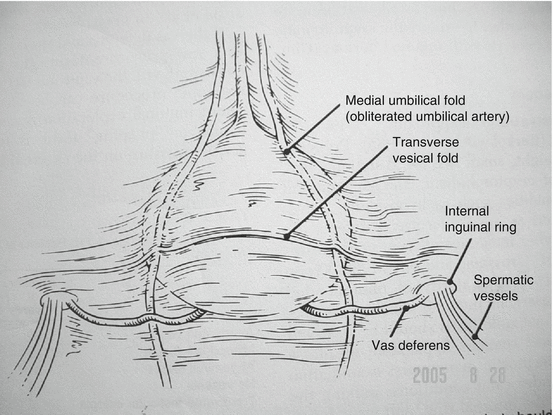
Once inside the peritoneal cavity, the normal side is examined first to reconfirm normal anatomy. The first landmark is the median umbilical fold (obliterated umbilical artery) on the anterior wall of the bladder. The vas deferens should cross over it from medial to lateral, running toward the internal ring. This is joined by the testicular vessels, which run parallel to the iliac vessels (see accompanying Video 18.1) (Fig. 18.2).


Fig. 18.2
The normal anatomy of the internal ring
The findings that can be seen at diagnostic laparoscopy include:
1.
Normal vas and vessels entering the canal with or without a patent process vaginalis. Occasionally a testicle can be seen peeping in from the internal ring (see accompanying Video 18.1).
2.
Intra-abdominal testis with normal vas and vessels with adequate mobility. This is usually assessed by seeing if the testis can reach the opposite internal ring (see accompanying Video 18.2).
3.
Intra-abdominal testis with short vessels and normal vas deferens (see accompanying Video 18.3).
4.
Vessels that become atretic before entering the internal ring. This represents an absent testicle. This is only true if the vessels can be seen and become atretic, not if the vas is not visualized alone (see accompanying Video 18.4).
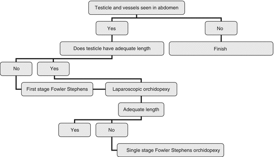
Once the diagnostic laparoscopy is performed, there are three treatment options if a testicle is seen: (1) a single-stage orchidopexy, (2) the first stage of a Fowler Stephens orchidopexy, or (3) a single-stage Fowler Stephens orchidopexy. Figure 18.3 proposes a management algorithm.


Fig. 18.3
Algorithm for the management of a patient with an intra-abdominal testicle
Single-Stage Laparoscopic Orchidopexy
Indication
An indication for this procedure is intra-abdominal or peeping testis with good vas and vessels that appear to have adequate length.
Port Position
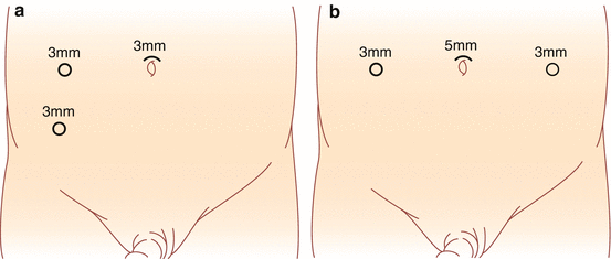
Following the placement of the camera port, two working ports are placed with local anesthetic under direct vision. The position of ports for a standard orchidopexy is shown in Fig. 18.4a, b).


Fig. 18.4


Port site position for a unilateral (a) or bilateral orchidopexy (b)
Stay updated, free articles. Join our Telegram channel

Full access? Get Clinical Tree


