Chapter 645 Hypopigmented Lesions
Albinism
Several types of congenital oculocutaneous albinism (OCA) consist of partial or complete failure of melanin production in the skin, hair, and eyes despite the presence of normal number, structure, and distribution of melanocytes. They may be divided into two major classes: those with abnormal protein function involved in the formation and transfer of melanin, and those with defects in melanosomes (Table 645-1). Tyrosinase is the copper-containing enzyme that catalyzes at multiple steps in melanin biosynthesis (Chapter 79.2). Tyrosinase-positive variants are characterized by darkening of the hair bulb on incubation with tyrosine.
Table 645-1 GENES ASSOCIATED WITH HYPOPIGMENTATION
| DISORDER | GENE DEFECT |
|---|---|
| Oculocutaneous albinism: | |
| OCA1 | Tyrosinase |
| OCA2 | P protein |
| OCA3 | TRP-1 |
| OCA4 | MATP |
| Hermansky-Pudlak: | |
| Type 1 | HPS-1 Mouse (pale ear) |
| Type 2 | HPS-2 b3A subunit of AP3 |
| Type 3 | HPS-3 Mouse (cocoa) |
| Type 4 | HPS-4 Mouse (light ear) |
| Type 5 | HPS-5 KIAA107 |
| Type 6 | HPS-6 Mouse (ruby eye) |
| Type 7 | HPS-7 DTNBP1 |
| Type 8 | HPS-8 Bloc153 |
| Chédiak-Higashi | CHS1/LYST |
| Piebaldism | C-KIT receptor |
| Heterozygous SLUG | |
| Waardenburg: | |
| Type 1 | Heterozygous PAX-3 |
| Type 2a | MITF |
| Type 2b | Chromosome 1p |
| Type 2c | Chromosome 8p23 |
| Type 2d | SNAIL |
| Type 2e | Sox 10 |
| Type 3 | Homozygous PAX-3 |
| Type 4 | SOX 10 |
| Endothelin 3 | |
| Endothelin B receptor | |
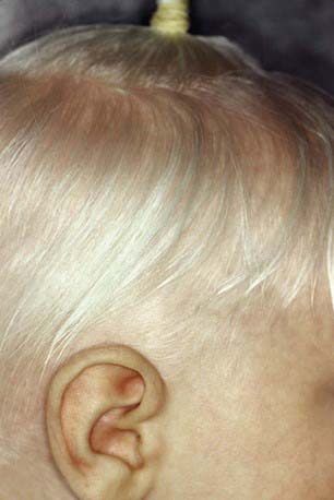
Oculocutaneous albinism type 1 (OCA1) is characterized by great reduction in or absence of tyrosinase activity. OCA1A, the most severe form, is characterized by a lack of visible pigment in hair, skin, and eyes (Fig. 645-1). This manifests as photophobia, nystagmus, defective visual acuity, white hair, and white skin. The irises are blue-gray in oblique light and prominent pink in reflected light. OCA1B, or yellow mutant albinism, manifests at birth as white hair, pink skin, and gray eyes. This type is particularly prevalent in Amish communities. Progressively the hair becomes yellow-red, the skin tans lightly on exposure to the sun, and the irises may accumulate some brown pigment, with a resultant improvement in visual acuity. Photophobia and nystagmus are present but mild. OCATS is a temperature-sensitive type of albinism. The abnormal tyrosinase has decreased activity at 35-37°C. Therefore, cooler regions of the body such as the limbs and head pigment to some degree, whereas other areas remain depigmented.
OCA4 is a rare OCA with clinical findings similar to those in OCA2.
Because of the absence of normal protection by adequate amounts of epidermal melanin, persons with albinism are predisposed to development of actinic keratoses and cutaneous carcinoma secondary to skin damage by ultraviolet light. Protective clothing and a broad-spectrum sunscreen preparation (Chapter 648) should be worn during exposure to sunlight.


