Hypofunction of the Ovaries

Chapter 580 Hypofunction of the Ovaries


Hypofunction of the ovaries can be either primary or central in etiology. It may be caused by congenital failure of development, postnatal destruction (primary or hypergonadotropic hypogonadism), or lack of central stimulation by the pituitary and/or hypothalamus (secondary or tertiary hypogonadotropic hypogonadism). Primary ovarian insufficiency (hypergonadotropic hypogonadism), which is also termed premature ovarian failure, is characterized by the arrest of normal ovarian function before the age of 40 yr. Mutations of certain genes could result in primary ovarian insufficiency. Hypofunction of the ovaries due to lack of central stimulation (hypogonadotropic hypogonadism) can be associated with other processes such as multiple pituitary hormone deficiencies and some chronic diseases. Table 580-1 details the etiologic classification of ovarian hypofunction.


580.1 Hypergonadotropic Hypogonadism in the Female (Primary Hypogonadism)


Diagnosis of hypergonadotropic hypogonadism before puberty is difficult. Except in the case of Turner syndrome, most affected patients have no prepubertal clinical manifestations.


Turner Syndrome


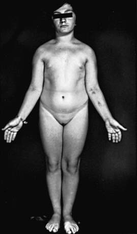
Turner described a syndrome consisting of sexual infantilism, webbed neck, and cubitus valgus in adult females (Chapter 76). Ullrich described an 8 yr old girl with short stature and many of the same phenotypic features. The term Ullrich-Turner syndrome is frequently used in Europe but rarely used in the USA. The condition is defined as the combination of the characteristic phenotypic features accompanied by complete or partial absence of the second X chromosome with or without mosaicism.


Pathogenesis


Half the patients with Turner syndrome have a 45,X chromosomal complement. About 15% of patients are mosaics for 45,X and a normal cell line (45,X/46,XX). Other mosaics with isochromosomes, 45,X/46,X,i(Xq); with rings, 45,X/46,X,r(X); or with fragments, 45,X/46fra, occur less often. Mosaicism is detected most commonly when more than one tissue is examined. The single X is of maternal origin in nearly 80% of 45,X patients. The mechanism of chromosome loss is unknown, and the risk for the syndrome does not increase with maternal age. The genes involved in the Turner phenotype are X-linked genes that escape inactivation. A major locus involved in the control of linear growth has been mapped within the pseudoautosomal region of the X chromosome (PAR1). SHOX, a homeobox-containing gene of 170 kb of DNA within the PAR1, is thought to be important for controlling growth in children with Turner syndrome, Leri-Weill syndrome, and rarely in patients having idiopathic short stature. Genes for the control of normal ovarian function are postulated to be on Xp and perhaps two “supergenes” on Xq.


Turner syndrome occurs in about 1/1,500-2,500 live born females. The frequency of the 45,X karyotype at conception is about 3.0%, but 99% of these are spontaneously aborted, accounting for 5-10% of all abortuses. Mosaicism (45,X/46,XX) occurs in a proportion higher than that seen with any other aneuploid state, but the mosaic Turner constitution is rare among the abortuses; these findings indicate preferential survival for mosaic forms.


The normal fetal ovary contains about 7 million oocytes, but these begin to disappear rapidly after the 5th mo of gestation. At birth, there are only 2 million (1 million active follicles); by menarche, there are 400,000-500,000; and at menopause, 10,000 remain. In the absence of 1 X chromosome, this process is accelerated, and nearly all oocytes are gone by 2 yr of age. In aborted 45,X fetuses, the number of primordial germ cells in the gonadal ridge appears to be normal, suggesting that the normal process is accelerated in patients with Turner syndrome. Eventually, the ovaries are described as “streaks” and consist only of connective tissue, with only a few germ cells present.


Clinical Manifestations


Many patients with Turner syndrome are recognizable at birth because of a characteristic edema of the dorsa of the hands and feet and loose skinfolds at the nape of the neck. Low birthweight and decreased length are common (Chapter 76). Clinical manifestations in childhood include webbing of the neck, a low posterior hairline, small mandible, prominent ears, epicanthal folds, high arched palate, a broad chest presenting the illusion of widely spaced nipples, cubitus valgus, and hyperconvex fingernails. The diagnosis is often first suspected at puberty when breast development fails to occur.


Short stature, the cardinal finding in virtually all girls with Turner syndrome, may be present with little in the way of other clinical manifestations. The linear growth deceleration begins in infancy and young childhood, gets progressively more pronounced in later childhood and adolescence, and results in significant adult short stature. Sexual maturation fails to occur at the expected age. Among untreated patients with Turner syndrome, the mean adult height is 143-144 cm in the USA and most of northern Europe, but 140 cm in Argentina and 147 cm in Scandinavia (Fig. 580-1). The height is well correlated with the midparental height (average of the parents’ heights). Specific growth curves for height have been developed for girls with Turner syndrome.


Associated cardiac defects are common. In the girls with Turner syndrome, life-threatening consequences of X-chromosome haploinsufficiency involve the cardiovascular system. There is a 4- to 5-fold increased rate of premature mortality secondary to congenital heart disease and premature coronary heart disease in adults with Turner syndrome. Clinically silent cardiac defects, mainly bicuspid aortic valve, but also ascending aortic dilation and partial anomalous pulmonary venous connections are present in patients with Turner syndrome. Regardless of the age, all patients with Turner syndrome at the time of diagnosis need comprehensive cardiovascular evaluation by a cardiologist specializing in congenital heart disease. Complete cardiologic evaluation, including echocardiography, reveals isolated nonstenotic bicuspid aortic valves in one third to one half of patients. In later life, bicuspid aortic valve disease can progress to dilatation of the aortic root. Less frequent defects include aortic coarctation (20%), aortic stenosis, mitral valve prolapse, and anomalous pulmonary venous drainage. In a study of 170/393 females with Turner syndrome in Denmark, 38% of patients with 45,X chromosomes had cardiovascular malformations compared with 11% of those with mosaic monosomy X; the most common were aortic valve abnormalities and aortic coarctation. Webbed neck in patients with or without recognized syndromes is associated with both flow-related and non–flow-related heart defects. Among patients with Turner syndrome, those with webbed neck have a much greater chance of having coarctation of the aorta than do those without webbed necks. Recent studies have suggested that in Turner syndrome there is a broader spectrum of cardiovascular abnormalities than previously recognized. Transthoracic echocardiogram in young girls is adequate if cardiac anatomy is clearly seen; otherwise magnetic resonance angiographic screening studies should be considered in asymptomatic individuals with Turner syndrome. During adolescence, and certainly before pregnancy is contemplated, repeat cardiac evaluation should be considered even in those without prior findings of cardiac abnormalities. Blood pressure should be routinely monitored even in the absence of cardiac or renal lesions and especially in those with suggestions of aortic root dilatation. Cardiac MRI is a valuable tool to detect and monitor aortic root dilation.


Renal ultrasound should be performed in all girls with Turner syndrome at diagnosis. One fourth to one third of patients have renal malformations on ultrasonographic examination (50% of those with 45,X karyotypes). The more serious defects include pelvic kidney, horseshoe kidney, double collecting system, complete absence of one kidney, and ureteropelvic junction obstruction. Some of the malformations may increase the risk of hypertension and urinary tract infection. Idiopathic hypertension is also common. Girls with Turner syndrome who had normal baseline renal ultrasound did not develop renal disease during a follow-up period averaging 6 yr.


When the ovaries were examined by ultrasonography, older studies found a significant decrease in percentage of detectable ovaries from infancy to later childhood. A subsequent report found no such age-related differences in a cross-sectional and longitudinal study conducted in Italy; 27-46% of patients had detectable ovaries at various ages; 76% of those with X mosaicism and 26% of those with 45,X karyotypes had detectable ovaries.


Sexual maturation usually fails to occur, but 10-20% of girls have spontaneous breast development, and a small percentage may have menstrual periods. Primary gonadal failure is associated with early onset of adrenarche (elevation in DHEA sulfate) but delayed pubarche (pubic hair development). Spontaneous pregnancies have been reported in menstruating patients with Turner syndrome. Premature menopause, increased risk of miscarriage, and offspring with increased risk of trisomy 21 have been reported in some of these women. A woman with a 45,X/46,X,r(X) karyotype treated with hormone replacement therapy had 3 pregnancies, resulting in a normal 46,XY male infant, a spontaneous abortion, and a healthy term female with Turner syndrome 45,X/46,Xr(X).


Antithyroid antibodies (thyroid peroxidase, and/or thyroglobulin antibodies) occur in 30-50% of patients. The prevalence increases with advancing age. Ten to 30% have autoimmune thyroid disease, with or without the presence of a goiter. Age-dependent abnormalities in carbohydrate metabolism characterized by abnormal glucose tolerance and insulin resistance and, only rarely, frank type 2 diabetes occur in patients with Turner syndrome. Impaired insulin secretion has been described in 45,X women. Cholesterol levels are elevated in adolescence, regardless of body mass index or karyotype.


Inflammatory bowel disease, both Crohn disease and ulcerative colitis; gastrointestinal bleeding due to abnormal mesenteric vasculature; and delayed gastric emptying time have all been reported. Screening for celiac disease is recommended by recent guidelines, since the risk of celiac disease is increased in Turner syndrome, with 4-6% of individuals affected. Although autoimmune diseases have been associated with Turner syndrome, the prevalence of type 1 diabetes with Turner syndrome is not very high.


Sternal malformations can be detected by lateral chest radiography. An increased carrying angle at the elbow is usually not clinically significant. Scoliosis occurs in about 10% of adolescent girls. Congenital hip dysplasia occurs more commonly than in the general population. Reported eye findings include anterior segment dysgenesis and keratoconus. Pigmented nevi become more prominent with age; melanocytic nevi are common. Essential hyperhidrosis, torus mandibularis, and alopecia areata occur rarely.


Recurrent bilateral otitis media develops in about 75% of patients. Sensorineural hearing deficits are common, and the frequency increases with age. Problems with gross and fine motor-sensory integration, failure to walk before 15 mo of age, and early language dysfunction often raise questions about developmental delay, but intelligence is normal in most patients. However, mental retardation does occur in patients with 45,X/46,X,r(X); the ring chromosome is unable to undergo inactivation and leads to 2 functional X chromosomes.


A special attention should be given to psychosocial development in girls with Turner syndrome. In general the behavior function is normal in girls with Turner syndrome, but they are at an increased risk for social isolation, immaturity, and anxiety. Other conditions such as dyslexia, nonverbal learning disability, and attention deficit disorder have been reported in girls with Turner syndrome. In adults, deficits in perceptual spatial skills are more common than they are in the general population. Some unconfirmed data suggest the existence of an imprinted X-linked locus that affects cognitive function such as verbal and higher-order executive function skills. These functions are apparently better when the X is paternal in origin.


The prevalence of mosaicism depends in large part on the techniques used for studying chromosomal patterns. The use of fluorescent in situ hybridization and reverse transcription–polymerase chain reaction (PCR) has increased the reported prevalence of mosaic patterns to as high as 60-74%.

Stay updated, free articles. Join our Telegram channel

Jun 18, 2016 | Posted by in PEDIATRICS | Comments Off on Hypofunction of the Ovaries

Full access? Get Clinical Tree

Get Clinical Tree app for offline access