Chapter 644 Hyperpigmented Lesions
Disorders of Pigment
Normal pigmentation requires migration of melanoblasts from the neural crest to the dermal-epidermal junction, enzymatic processes to form pigment, structural components to contain the pigment (melanosomes), and transfer of pigment to the surrounding keratinocytes. Increased skin color may be generalized or localized and may result from various defects in any of these requirements. Some of these aberrations are a manifestation of systemic disease, others represent generalized or focal developmental or genetic defects, and still others may be nonspecific and the result of cutaneous inflammation.
Ephelides (Freckles)
Ephelides are light or dark brown macules usually <3 mm in diameter, with a poorly defined margin, that occur in sun-exposed areas such as the face, upper back, arms, and hands. They are induced by exposure to sun, particularly during the summer, and may fade or disappear during the winter. They are more common in redheads and fair-haired individuals and first appear in the preschool years. Histologically, they are marked by increased melanin pigment in epidermal basal cells, which have more numerous and larger dendritic processes than the melanocytes of the surrounding paler skin. The lack of melanocytic proliferation or elongation of epidermal rete ridges distinguishes them from lentigines. Freckles have been identified as a risk factor for melanoma independent of melanocytic nevi.
Lentigines
Lentigines, often mistaken for freckles or junctional nevi, are small (<3 cm), round, dark brown macules that can appear anywhere on the body. They are unrelated to sun exposure and remain permanently. Histologically, they have elongated, club-shaped, epidermal rete ridges with increased numbers of melanocytes and dense epidermal deposits of melanin. No nests of melanocytes are found. The lesions are benign and, when few, may be viewed as a normal occurrence and are seen most commonly on the lower lip.
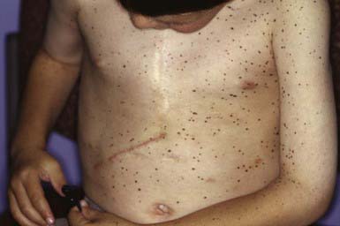
Lentiginosis profusa involves innumerable small, pigmented macules that are present at birth or appear during childhood. There are no associated abnormalities, and mucous membranes are spared. LAMB syndrome (Carney complex), a multiple endocrine neoplasia syndrome, consists of lentigines of the face and vulva, atrial myxoma, mucocutaneous myxomas, and blue nevi (type 1, PRKAR1 gene; type 2, gene map locus 2p16-gene as yet to be identified). The Carney complex variant is associated with distal arthrogryposis (MYH8 gene). The multiple lentigines (LEOPARD) syndrome is an autosomal dominant entity consisting of a generalized, symmetric distribution of lentigines (Fig. 644-1) in association with electrocardiogram abnormalities, ocular hypertelorism, pulmonary stenosis, abnormal genitals (cryptorchidism, hypogonadism, hypospadias), growth retardation, and sensorineural deafness (type 1, PTPN11 gene; type 2, RAF1 gene). Other features include hypertrophic obstructive cardiomyopathy and pectus excavatum or carinatum.

Figure 644-1 Multiple lentigines in LEOPARD (lentigines in association with electrocardiogram abnormalities, ocular hypertelorism, pulmonary stenosis, abnormal genitals [cryptorchidism, hypogonadism, hypospadias], growth retardation, and sensorineural deafness) syndrome.

Stay updated, free articles. Join our Telegram channel

Full access? Get Clinical Tree

