Human Metapneumovirus

Chapter 253 Human Metapneumovirus


Human metapneumovirus (HMPV), a respiratory virus identified in 2001, is emerging as one of the most common causes of serious lower respiratory tract illness in children throughout the world.


Etiology


HMPV is an enveloped, single-stranded nonsegmented negative-sense RNA genome of the Paramyxoviridae family, which is divided into 2 subfamilies, Pneumovirinae and Paramyxovirinae. The Pneumovirinae subfamily includes the 2 genera Metapneumovirus and Pneumovirus, which includes respiratory syncytial virus (RSV). HMPV and the avian pneumoviruses (APVs) are highly related and are separated into the separate genus Metapneumovirus because the gene order in the nonsegmented genome is slightly altered and APVs/HMPVs lack the genes for 2 nonstructural proteins, NS1 and NS2, that are encoded at the 3′ end of RSV genomes. These proteins are thought to counteract host type I interferons. The absence of NS1/NS2 in the metapneumoviruses may contribute to decreased pathogenicity of HMPV relative to wild-type RSV strains.


Full-length sequences of a number of HMPV genomes have been determined. The genome is predicted to encode 9 proteins in the order 3′-N-P-M-F-M2-(orf1 and 2)-SH-G-L-5′. The genome also contains noncoding 3′ leader, 5′ trailer, and intergenic regions, consistent with the organization of most paramyxoviruses, with a viral promoter contained in the 3′ end of the genome. The F (fusion), G (glycosylated), and SH (short hydrophobic) proteins are integral membrane proteins on the surfaces of infected cells and virion particles. The F protein is a classic type I integral membrane viral fusion protein that contains 2 heptad repeats in the extracellular domain that facilitate membrane fusion. There is a predicted protein cleavage site near a hydrophobic fusion peptide that likely is cleaved by an extracellular protease, activating the F protein for fusion. The predicted attachment (G) protein of HMPV exhibits the basic features of a glycosylated type II mucin-like protein. The HMPV G protein differs from the RSV G protein in that it lacks a cysteine noose structure. This protein may inhibit innate immune responses. The internal proteins of the virus appear similar in function to those of other paramyxoviruses.


Epidemiology


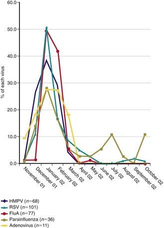
HMPV outbreaks occur in annual epidemics during late winter and early spring in temperate climates, often overlapping with the second half of the annual RSV epidemic (Fig. 253-1). Sporadic infection does occur year round. The usual period of viral shedding is likely to be several weeks after primary infection in infants. The incubation period is 3-5 days. Humans are the only source of virus. Transmission is thought to occur by close or direct contact with contaminated secretions involving large-particle aerosols, droplets, or contaminated surfaces. Nosocomial infections have been reported, and contact isolation with excellent handwashing for health care providers is indicated in medical settings. This virus affects the elderly, immunocompromised patients, and patients with reactive airways disease more severely than otherwise healthy individuals.


Jun 18, 2016 | Posted by in PEDIATRICS | Comments Off on Human Metapneumovirus

Full access? Get Clinical Tree

Get Clinical Tree app for offline access