High Output Heart Failure
Eva Ilse Rubio, MD
DIFFERENTIAL DIAGNOSIS
Common
Intracardiac Shunt
Vein of Galen Aneurysmal Malformation
Anemias
Less Common
Vascular Malformations
Hemangioendothelioma
Teratoma
Rare but Important
Parkes-Weber
Chorioangioma
ESSENTIAL INFORMATION
Key Differential Diagnosis Issues
Age of patient: Newborn, toddler, child, or adolescent?
Prior medical history
Underlying congenital heart defect or aortic anomaly
Infectious history or postinfectious syndrome
Chronic medical conditions or therapy
Intracardiac vs. extracardiac cause?
Helpful Clues for Common Diagnoses
Intracardiac Shunt
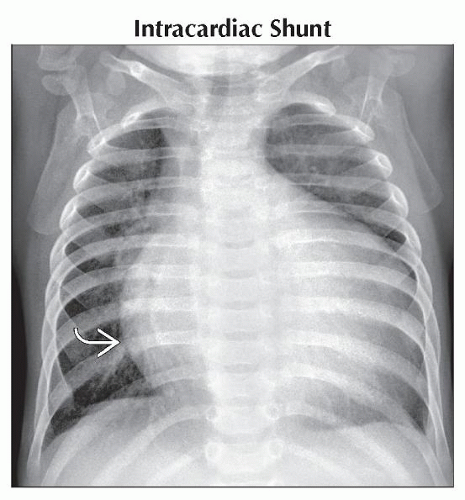
Ventricular septal defect
Most common cardiac anomaly in general population
Radiograph findings: Hyperexpanded lungs, large round heart, plump pulmonary vessels most conspicuous at hila
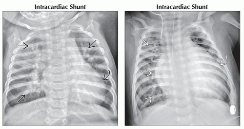
Atrioventricular septal defect
Commonly associated with Down syndrome
Radiograph findings: Hyperexpanded lungs, large round heart, plump pulmonary vessels most conspicuous at hila
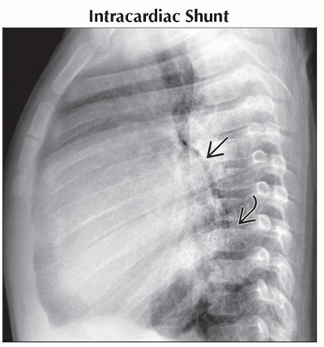
Anomalous coronary artery
Most commonly, left coronary artery arises from pulmonary artery origin
Preferential flow away from myocardial muscular bed into pulmonary circulation results in ventricular ischemia
Imaging features: Marked cardiomegaly (CM); left ventricular and atrial enlargement conspicuous on lateral view
Vein of Galen Aneurysmal Malformation
Congenital arteriovenous fistulous communicate between midline intracranial arteries and vein of Galen or other fetal venous structures
Poor prognosis
Choroidal type
Fetal hydropic changes
Congestive heart failure with intracranial bruit
Intracranial findings
Large midline vascular anomaly
Hydrocephalus
Encephalomalacia
Anemias
Sickle cell disease
Longstanding anemia leads to global CM
Physiologic contributions: High output of anemia, poor oxygen delivery to coronary arteries, pulmonary arterial hypertension
More common in older children, adolescents
β-thalassemia
Severe anemia leads to global CM
Physiologic contributions: High output anemia, elevated iron levels/deposition from transfusions
May manifest earlier in childhood
Helpful Clues for Less Common Diagnoses
Vascular Malformations
Typical locations: Head, neck, extremities, liver
Classification of vascular anomalies (these lesions grow commensurate with child)
High-flow vascular lesions (arteriovenous malformations and arteriovenous fistulae)
Low-flow vascular lesions (venous, lymphatic, venolymphatic)
Hemangioendothelioma
a.k.a. infantile hepatic hemangioma
Hypervascular liver mass seen in infants
May have cutaneous hemangiomas, especially multiple
Imaging
May be multiple rounded lesions or single dominant lesion
US: Hypoechoic
CT: Round, enhancing lesions; may enhance from periphery to center
MR: Bright on T2, isointense/dark on T1
Cardiovascular sequelae/manifestations
Significant arteriovenous shunting may lead to high output failure
Aorta may be diminutive distal to lesion
Disseminated intravascular coagulopathy may occur
Teratoma
Germ cell tumor presumably arising from multipotential cells
Typical locations
Sacrococcygeal region, arising from Hensen node
Neck
Oropharynx
Abdomen
Retroperitoneum
More common in females
Findings associated with increased risk for congestive heart failure
Larger lesions
Significant solid tissue component
Large feeding vessels/robust internal vascularity
Intralesional hemorrhage
Placentomegaly
Hydropic changes in fetus
Sacrococcygeal teratoma (SCGT)
Classified into types 1-4 based on extrapelvic vs. intrapelvic components
SCGT findings associated with heart failure: Aortic velocity > 60 cm/sec; IVC diameter > 4.1 mm (21-28 weeks gestation); reversed hypogastric flow
Cervical teratomas
Differential consideration is lymphatic malformation if multicystic
Other considerations: Airway management, intracranial involvement
Helpful Clues for Rare Diagnoses
Parkes-Weber
Rare extremity soft tissue overgrowth syndrome due to vascular anomaly
Limb overgrowth
Combined vascular malformation; capillary malformation, high-flow AVMs ± lymphatic malformation
May result in high output heart failure
Chorioangioma
Benign, rare placental tumor
Consists of small caliber vascular structures and stroma
Often seen at base of umbilical cord, with vascular supply arising near or from umbilical vessels
Potential fetal sequelae
Hydropic changes
Growth restriction
Image Gallery
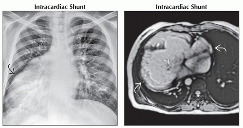
 (Left) AP radiograph shows a patient with cardiomegaly
 due to severe complex congenital heart disease, including situs inversus, transposition of the great vessels, and large atrial/ventricular septal defects. (Right) Axial T2WI MR of the same patient shows global enlargement of all 4 chambers of the heart due to severe complex congenital heart disease, including situs inversus, transposition of the great vessels, and large atrial/ventricular septal defects. (Right) Axial T2WI MR of the same patient shows global enlargement of all 4 chambers of the heart  . The large ventricular septal defect is depicted . The large ventricular septal defect is depicted  . Cine imaging demonstrated globally depressed ventricular systolic function. . Cine imaging demonstrated globally depressed ventricular systolic function.Stay updated, free articles. Join our Telegram channel
Full access? Get Clinical Tree
 Get Clinical Tree app for offline access

|


