Heart Failure

Chapter 436 Heart Failure


Heart failure occurs when the heart cannot deliver adequate cardiac output to meet the metabolic needs of the body. In the early stages of heart failure, various compensatory mechanisms are evoked to maintain normal metabolic function. When these mechanisms become ineffective, increasingly severe clinical manifestations result (Chapter 64).


Pathophysiology


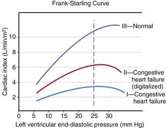
The heart can be viewed as a pump with an output proportional to its filling volume and inversely proportional to the resistance against which it pumps. As ventricular end-diastolic volume increases, a healthy heart increases cardiac output until a maximum is reached and cardiac output can no longer be augmented (the Frank-Starling principle; Fig. 436-1). The increased stroke volume obtained in this manner is due to stretching of myocardial fibers, but it also results in increased wall tension, which elevates myocardial oxygen consumption. Hearts working under various types of stress function along different Frank-Starling curves. Cardiac muscle with compromised intrinsic contractility requires a greater degree of dilatation to produce increased stroke volume and does not achieve the same maximal cardiac output as normal myocardium does. If a cardiac chamber is already dilated because of a lesion causing increased preload (e.g., a left-to-right shunt or valvular insufficiency), there is little room for further dilatation as a means of augmenting cardiac output. The presence of lesions that result in increased afterload to the ventricle (aortic or pulmonic stenosis, coarctation of the aorta) decreases cardiac performance, thereby resulting in a depressed Frank-Starling relationship. The ability of an immature heart to increase cardiac output in response to increased preload is less than that of a mature heart. Thus, premature infants are more compromised by a left-to-right shunt than full-term infants are.


Systemic oxygen transport is calculated as the product of cardiac output and systemic oxygen content. Cardiac output can be calculated as the product of heart rate and stroke volume. The primary determinants of stroke volume are the afterload (pressure work), preload (volume work), and contractility (intrinsic myocardial function). Abnormalities in heart rate can also compromise cardiac output; for example, tachyarrhythmias shorten the diastolic time interval for ventricular filling. Alterations in the oxygen-carrying capacity of blood (e.g., anemia or hypoxemia) also lead to a decrease in systemic oxygen transport and, if compensatory mechanisms are inadequate, can result in decreased delivery of substrate to tissues.


In some cases of heart failure, cardiac output is normal or increased, yet because of decreased systemic oxygen content (secondary to anemia) or increased oxygen demands (secondary to hyperventilation, hyperthyroidism, or hypermetabolism), an inadequate amount of oxygen is delivered to meet the body’s needs. This condition, high-output failure, results in the development of signs and symptoms of heart failure when there is no basic abnormality in myocardial function and cardiac output is greater than normal. It is also seen with large systemic arteriovenous fistulas. These conditions reduce peripheral vascular resistance and cardiac afterload and increase myocardial contractility. Heart “failure” results when the demand for cardiac output exceeds the ability of the heart to respond. Chronic severe high-output failure may eventually result in a decrease in myocardial performance as the metabolic requirements of the myocardium are not met.


There are multiple systemic compensatory mechanisms used by the body to adapt to chronic heart failure. Some are mediated at the molecular/cellular level, such as upregulation or downregulation of various metabolic pathway components leading to changes in efficiency of oxygen and other substrate utilization. Others are mediated by neurohormones such as the renin-angiotensin system and the sympathoadrenal axis. One of the principal mechanisms for increasing cardiac output is an increase in sympathetic tone secondary to increased secretion of circulating epinephrine by the adrenals and increased release of norepinephrine at the neuromuscular junction. The initial beneficial effects of sympathetic stimulation include an increase in heart rate and myocardial contractility, mediated by these hormones’ action on cardiac β-adrenergic receptors, increasing cardiac output. These hormones also cause vasoconstriction, mediated by their action on peripheral arterial α-adrenergic receptors. Some vascular beds may constrict more readily than others, so that blood flow is redistributed from the cutaneous, visceral, and renal beds to the heart and brain. Whereas these acute effects are beneficial, chronically increased sympathetic stimulation can have deleterious effects, including hypermetabolism, increased afterload, arrhythmogenesis, and increased myocardial oxygen requirements. Peripheral vasoconstriction can result in decreased renal, hepatic, and gastrointestinal tract function. Chronic exposure to circulating catecholamines leads to a decrease in the number of cardiac β-adrenergic receptors (downregulation) and also causes direct myocardial cell damage. Thus, therapeutic agents for heart failure are directed at restoring balance to these neuroendocrine systems. Single nucleotide differences (known as polymorphisms [SNPs]) in the genes encoding proteins involved in sympathetic signaling can alter a patient’s response to medical therapy and may predict risk of worsening heart failure, hospitalization, or death. These pharmacogenomic studies may allow us to tailor our therapies to the individual patient, based on their genetic makeup.


Clinical Manifestations


The clinical manifestations of heart failure depend on the degree of the child’s cardiac reserve. A critically ill infant or child who has exhausted the compensatory mechanisms to the point that cardiac output is no longer sufficient to meet the basal metabolic needs of the body will be symptomatic at rest. Other patients may be comfortable when quiet but are incapable of increasing cardiac output in response to even mild activity without experiencing significant symptoms. Conversely, it may take rather vigorous exercise to compromise cardiac function in children who have less severe heart disease. A thorough history is extremely important in making the diagnosis of heart failure and in evaluating the possible causes. Parents who observe their child on a daily basis may not recognize subtle changes that have occurred over the course of days or weeks. Gradually worsening perfusion or increasing respiratory effort may not be recognized as an abnormal finding. Edema may be passed off as normal weight gain, and exercise intolerance as lack of interest in an activity. The history of a young infant should also focus on feeding (Chapter 416). An infant with heart failure often takes less volume per feeding, becomes dyspneic while sucking, and may perspire profusely. Eliciting a history of fatigue in an older child requires detailed questions about activity level and its course over several months.


In children, the signs and symptoms of heart failure may be similar to those in adults and include fatigue, effort intolerance, anorexia, dyspnea, and cough. Many children, however, especially adolescents, may have primarily abdominal symptoms (abdominal pain, nausea, anorexia) and a surprising lack of respiratory complaints. Attention to the cardiovascular system may come only after an abdominal roentgenogram unexpectedly catches the lower end of an enlarged heart. The elevation in systemic venous pressure may be gauged by clinical assessment of jugular venous pressure and liver enlargement. Orthopnea and basilar rales are variably present; edema is usually discernible in dependent portions of the body, or anasarca may be present. Cardiomegaly is invariably noted. A gallop rhythm is common; when ventricular dilatation is advanced, the holosystolic murmur of mitral or tricuspid valve regurgitation may be heard.


In infants, heart failure may be difficult to distinguish from other causes of respiratory distress. Prominent manifestations include tachypnea, feeding difficulties, poor weight gain, excessive perspiration, irritability, weak cry, and noisy, labored respirations with intercostal and subcostal retractions, as well as flaring of the alae nasi. The signs of cardiac-induced pulmonary congestion may be indistinguishable from those of bronchiolitis; wheezing is often a more prominent finding in young infants with heart failure than rales. Pneumonitis with or without atelectasis is common, especially in the right middle and lower lobes, due to bronchial compression by the enlarged heart. Hepatomegaly usually occurs, and cardiomegaly is invariably present. In spite of pronounced tachycardia, a gallop rhythm can frequently be recognized. The other auscultatory signs are those produced by the underlying cardiac lesion. Clinical assessment of jugular venous pressure in infants may be difficult because of the shortness of the neck and the difficulty of observing a relaxed state; palpation of an enlarged liver is a more reliable sign. Edema may be generalized and usually involves the eyelids as well as the sacrum and less often the legs and feet. The differential diagnosis is age dependent (Table 436-1).


Diagnosis


X-rays of the chest show cardiac enlargement. Pulmonary vascularity is variable and depends on the cause of the heart failure. Infants and children with large left-to-right shunts have exaggeration of the pulmonary arterial vessels to the periphery of the lung fields, whereas patients with cardiomyopathy may have a relatively normal pulmonary vascular bed early in the course of disease. Fluffy perihilar pulmonary markings suggestive of venous congestion and acute pulmonary edema are seen only with more severe degrees of heart failure. Cardiac enlargement is often noted as an unexpected finding on a chest roentgenogram performed to evaluate for a possible pulmonary infection, bronchiolitis, or asthma.


Chamber hypertrophy noted by electrocardiography may be helpful in assessing the cause of heart failure but does not establish the diagnosis. In cardiomyopathies, left or right ventricular ischemic changes may correlate with other noninvasive parameters of ventricular function. Low-voltage QRS morphologic characteristics with ST-T wave abnormalities may also suggest myocardial inflammatory disease but can be seen with pericarditis as well. The electrocardiogram is the best tool for evaluating rhythm disorders as a potential cause of heart failure, especially tachyarrhythmias.


Echocardiography is the standard technique for assessing ventricular function. The most commonly used parameter in children is fractional shortening (a single dimensional variable), determined as the difference between end-systolic and end-diastolic diameter divided by end-diastolic diameter. Normal fractional shortening is between approximately 28% and 42%. In adults, the most commonly used parameter is ejection fraction (which uses two-dimensional data to calculate a three-dimensional volume) and the normal range is 55-65%. In children with right ventricular enlargement or other cardiac pathology resulting in flattening of the interventricular septum, ejection fraction is used since fractional shortening measured in the standard echocardiographic short-axis view will not be accurate. Doppler studies can also be used to estimate cardiac output. Doppler tissue imaging is a new technique which can assess not only cardiac function, but wall motion abnormalities that can interfere with normal synchronous cardiac contraction. Magnetic resonance angiography (MRA) is also very useful in quantifying left and right ventricular function, volume and mass. If valvar regurgitation is present, MRA can quantify the regurgitant fraction.

Stay updated, free articles. Join our Telegram channel

Jun 18, 2016 | Posted by in PEDIATRICS | Comments Off on Heart Failure

Full access? Get Clinical Tree

Get Clinical Tree app for offline access