Fig. 1
(Dinh 2009) Clinical photo demonstrating the subcutaneous location of the metacarpal head in a dorsal metacarpophalangeal dislocation. Note the position of the radial digital nerve which can often be displaced volarly just under the skin making it prone to injury with this approach
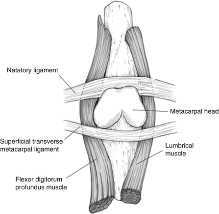
Fig. 2
(Dinh 2009) Schematic representation of common obstacles to reductions. The metacarpal head is often trapped between the flexor tendons ulnarly and lumbricals radially. The natatory ligament lies distally, while the superficial transverse metacarpal ligament is positioned proximally
Dislocations of the MCP joint can be divided into complex or simple. In simple dislocations, the volar plate remains volar or just distal to the articular surface of the metacarpal head. The term complex dislocation has been widely applied to those injuries in which closed reduction is not possible. Multiple authors have described this complex lesion and the failure of closed reduction (Baldwin et al. 1967; Burman 1953; Hunt et al. 1967; McLaughlin 1965; Milch 1965; Murphy and Stark 1967). This is most often due to the trapped volar plate that becomes positioned dorsal to the metacarpal head; however, several structures can sesamoids ultimately be responsible for an inability to reduce the joint. The natatory ligaments, palmar fascia, superficial transverse metacarpal ligament, lumbricals, and the flexor tendons have all been reported as possible obstacles to joint reduction (Kaplan 1957; Beatty et al. 1990; Plancher 2004). The flexor tendons and lumbricals are often found locked dorsally behind the displaced metacarpal head. Often the lumbrical can be found on the radial side of the metacarpal and the flexor tendons on the ulnar side contributing to a noose-like tightening made worse with traction. The A1 pulley is often still attached to the volar plate and pulls the flexor tendons dorsal to the metacarpal head.
Assessment of Metacarpophalangeal Dislocations
Examination of a child’s hand poses many challenges that are not present in the adult patient. Pain, stranger anxiety, and lack of understanding all contribute to the difficulty of the hand examination in a child. Clinically, the finger appears shortened, supinated, and angulated in an ulnar direction when an MCP dislocation is present. The skin may appear puckered volarly where the metacarpal head has buttonholed through the volar joint capsule. Careful examination of the palm skin is necessary to ensure there is no evidence of an open injury. Absent and painful attempts at MCP range of motion are often present. The metacarpophalangeal joint may exhibit several different positions. The joint may appear hyperextended as it does in a simple dislocation (or subluxation), which often represents a tear of the volar plate without dorsal displacement behind the metacarpal head. Alternatively, the proximal phalanx may be lying dorsal and parallel to the metacarpal. This is classically the complex dislocation in which closed reduction is not possible.
Swelling, bruising, and pain are all frequent findings with both types of dislocations (Fig. 3). Careful examination of the neurovascular status distal to the injury is equally important. The clinician should assess for adequate blood flow by comparing the color, temperature, capillary refill, and skin turgor of the finger to an uninjured digit. Palpation of the palm may reveal a bony prominence representing the metacarpal head. Decreased sensation to light touch and diminished two-point discrimination suggest pressure on the digital nerves which may be tented volarly over the metacarpal head. If a pediatric patient is unable to participate in two-point discriminatory testing, the wrinkle test may be performed by submerging the hand in water for several minutes if nerve dysfunction is suspected. Absence of wrinkling is indicative of nerve injury. A thorough sensory exam may be limited by pain in the finger, but in operative cases, once the patient is asleep, rubbing a sterile plastic marking pen back and forth, from the radial to the ulnar side of the finger, should be met with some resistance. If not, and the pen glides back and forth over the fingertip as if over a smooth surface on the involved finger in comparison to the other fingers, the examiner can conclude that the sympathetic innervation to smooth muscles responsible for skin turgor has been disrupted and a nerve injury to that finger has occurred.
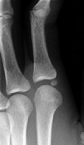
Fig. 3
(Plancher 2004) Radiograph demonstrating an entrapped sesamoid in the metacarpophalangeal joint
Imaging
A complete radiographic series, including posterior-anterior (PA) and lateral views (Fig. 4), in combination with a detailed clinical examination, should always be obtained on initial contact with the patient. The dislocation can often be difficult to interpret with a PA radiograph alone. With this view, the MCP joint may appear narrowed, distracted, angulated, or even normal in appearance (Light and Ogden 1988). Lateral plain films can often be obscured by the overlapping metacarpals. A line drawn straight down the shaft of the proximal phalanx should always intersect the metacarpal head. Failure of the line to intersect the metacarpal head suggests subluxation or dislocation (Campbell 1990). On the lateral view, the proximal phalanx lying parallel to the metacarpal shaft indicates a complex dislocation, while a simple dislocation is inferred if the proximal phalanx is oriented 90° to the metacarpal. In a child older than 10 years of age, sesamoids that have undergone ossification may also be observed in the joint (Green and Terry 1973; Campbell 1990).
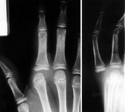
Fig. 4
(Plancher 2004) Radiograph of a complex metacarpophalangeal dislocation. Note the almost parallel relationship of the metacarpal and proximal phalanx (need a skeletally immature patient here)
More advanced imaging including computed tomography (CT) and magnetic resonance imaging (MRI) is rarely necessary for the diagnosis of these injuries. If the dislocation is associated with a fracture of the proximal phalanx or metacarpal head, a CT may permit better visualization of the articular anatomy, especially if an intra-articular fracture is suspected. These tests should only be reserved for rare cases in which a complete understanding of the bony anatomy is not possible with plain radiographs alone.
Classification
Dislocations are described by the direction of the distal part and the ability to achieve closed reduction. The term simple routinely refers to those dislocations in which closed reduction is successful, while complex dislocations require open reduction. A complex dislocation represents bayoneting of the proximal phalanx on the metacarpal. Alternatively, a simple joint dislocation is demonstrated by joint subluxation or perching of the proximal phalanx on the metacarpal. Subluxation of the joint can easily be converted to a complete (and complex) dislocation by exaggeration of the hyperextension or an incorrect reduction maneuver.
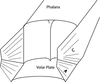
Fig. 5
Diagram depicting the three sided box formed by the accessory collateral ligaments and the volar plate. C: proper collateral ligament A: accessory collateral ligament (credit: Rey Ramirez)
Outcome Tools
Successful management of these injuries in the pediatric population should include full restoration of strength and range of motion. Assessments of grip strength, pinch strength, and range of motion for all the joints of the finger are important to measure and document. However, no outcome tool exists that is specific for pediatric hand dislocations.
Treatment Options for Dorsal Dislocation of the Metacarpophalangeal Joint
Nonoperative Management
Indications/Contraindications
An attempt at closed reduction should be made prior to any surgical intervention. While the complete dislocation is often irreducible by closed means, the clinician may attempt closed reduction of the joint to preclude the need for surgery. Multiple attempts at closed reduction should not be performed as it can lead to damage of the growth plate or rarely premature growth arrest of the metacarpal physis (Green and Terry 1973) (Table 1).
Table 1
Metacarpophalangeal joint dislocations | |
|---|---|
Nonoperative treatment | |
Indications | Contraindications |
Successful closed reduction | Closed reduction unsuccessful |
Chronic dislocation (trial of closed reduction should be attempted) | |
Fracture dislocation requiring open treatment | |
Open dislocation | |
Techniques
Appropriate analgesia is important prior to any attempt at a closed reduction. A digital nerve block proximal to the metacarpophalangeal joint with appropriate blocking of the dorsal and volar nerves is preferred. Attempts at closed reduction should focus on the type of dislocation. Simple dislocations often will reduce with gentle traction and volar translation. The translation is often much more important than the traction. If a complete dislocation is suspected, MCP joint hyperextension with distal and palmar translation of the proximal phalanx should be attempted. Excessive traction may result in a “noose-like” mechanism of soft tissue surrounding the metacarpal head. Hyperextension or traction alone may also convert an otherwise simple dislocation into a complex lesion (McLaughlin 1965; Campbell 1990). Additionally described techniques involve injection of the joint with a local anesthetic or saline in an attempt to deliver the palmar plate volar to the metacarpal head (O’Brien 1991).
If closed reduction is successful, the joint should be immobilized in a dorsal blocking splint with the joint in slight flexion and the wrist in either neutral or slight extension. Placement of the wrist in flexion may contribute to MCP extension as the extensor tendons are placed under greater tension. A trial of 3–4 weeks of immobilization should be followed with protected range of motion. Buddy taping is a useful tool to prevent additional instability while focusing on restoration of joint motion.
Outcomes
Long-term outcome studies of this rare injury have not been reported in the literature. While the adage “all things pediatric do well” is not always true, recognition of the injury pattern, appropriate reduction, and subsequent immobilization are all keys to achieving a good outcome. Formal hand therapy is rarely necessary as age-appropriate play activities usually result in full and stable range of motion with appropriate treatment. Long-term complications of these injuries are infrequent and are most often the result of a delay in diagnosis or treatment. While good outcomes have been reported in the treatment of missed dislocations of greater than 3 months, results tend to deteriorate as time to treatment increases (Barenfeld and Weseley 1972; Murphy and Stark 1967).
Operative Management
Indications/Contraindications
See Table 2.
Table 2
Metacarpophalangeal joint dislocations | |
|---|---|
Operative treatment | |
Indications | Contraindications |
Closed reduction unsuccessful | Closed reduction successful |
Chronic dislocation (trial of closed reduction should be attempted) | |
Fracture dislocation requiring open treatment | |
Open dislocations | |
Surgical Procedure
Preoperative Planning
Prior to surgical reduction of the joint, the clinician should obtain appropriate radiographic views and document a detailed physical exam. A complete understanding of the offending structures and challenges of each approach is necessary (Table 3).
Table 3
Open reduction for metacarpophalangeal joint dislocation |
|---|
Preoperative planning |
OR table: radiolucent hand table |
Position: supine |
Fluoroscopy: brought in from end of hand table |
Equipment: standard hand surgery set, Kirschner wire set |
Tourniquet: nonsterile |
Surgical Approach and Technique
Both dorsal and volar approaches have been described for the irreducible dorsal MCP dislocation. The volar approach provides superior visualization of the metacarpal head and additional structures (other than the volar plate) that may be blocking the reduction (Gilbert 1985; Kaplan 1957; Light and Ogden 1988; McLaughlin 1965; Green and Terry 1973; Barenfeld and Weseley 1972). If a volar approach is utilized, extreme care must be taken when making the skin incision to avoid injury to a digital nerve, in the case of the index finger, the radial digital nerve. An oblique Bruner-type incision should be centered over the metacarpal head. In the case of an open injury, the laceration is frequently extended to allow exposure of the injury. Iatrogenic nerve injury has been reported as the digital nerve is immediately beneath the skin as it becomes tented volarly by the displaced metacarpal head (Becton et al. 1975; Green and Terry 1973). After identification of the digital nerves and blunt soft tissue dissection, the surgeon may incise the A1 pulley. With attachments on either side of the volar plate, transection of the pulley may relax the tissue enough to facilitate reduction. Prior to reduction, a careful examination of the articular surface should be performed looking for loose bodies or osteochondral fragments. The metacarpal head should be delivered dorsally with retraction of the flexor tendons and lumbrical. A skin hook may be used to retrieve the dorsally displaced volar plate. If reduction can still not be accomplished, some surgeons advocate a longitudinal incision in the volar plate. This functions to relax the accessory collateral ligaments that also share an attachment on the volar plate. More chronic dislocations may require release of the ulnar collateral ligament to facilitate joint reduction. Once the joint is reduced, the finger should be taken through a gentle range of motion to determine a stable arc of motion. Typically no additional closure other than the skin is needed (Table 4).
Table 4
Volar approach of dorsal dislocation of the metacarpophalangeal joint |
|---|
Surgical steps |
Bruner incision centered on MCP head |
Blunt dissection to identify digital nerves |
Incise the A1 pulley |
Extract volar plate from MCP joint |
Examine joint for articular damage |
Reduce joint |
Determine stable arc for range of motion |
Close skin |
Several authors prefer a dorsal approach as it has the advantages of avoiding potential damage to the digital nerve and easier visualization of the dorsally dislocated volar plate (Becton et al. 1975; Bohart et al. 1982; McLaughlin 1965; Hunt et al. 1967). A curvilinear midline longitudinal incision is made centered over the metacarpophalangeal joint. The extensor tendon is identified and longitudinally split before a capsulotomy is made into the joint. Successful reduction from the dorsal approach often requires a longitudinal incision in the volar plate. Regardless of which approach is preferred, hardware including Kirschner wires is rarely necessary and may cause additional damage to the immature growth plate (Ogden 1982; Baldwin et al. 1967). A dual approach incorporating both volar and dorsal approaches may be needed for chronic dislocations with a delay in diagnosis (Murphy and Stark 1967; Barenfeld and Weseley 1972; Table 5).
Table 5
Open reduction metacarpophalangeal joint postoperative protocol |
|---|
Dorsal blocking splint to fingertips for dorsal dislocation |
Length of immobilization: 2 weeks followed by active range of motion with a dorsal blocking splint for 2 weeks |
Rehab protocol: buddy taping for 1–2 weeks. Encourage early range of motion within the stable arc |
Return to sport when range of motion returns and pain is minimal |
For very small children who require splinting, consider placing a thermoplast splint directly on the skin to ensure joint position and then overwrap it with cast material |
Preferred Treatment
If a complex dislocation is present, a volar approach is preferred because all offending structures can be identified. Reduction of the volar plate can often be accomplished with release of the A1 pulley and a translation maneuver. If a relaxing incision is used, repair of the volar plate can easily be performed. While the dorsal approach can be successful, one disadvantage is the inability to release the volar structures, such as the A1 pulley. Additional structures, such as the palmar aponeurosis, may also prevent the metacarpal head from reducing (Kaplan 1957). Release of these structures requires a volar approach. Additionally, suture repair of the volar plate incision is not possible from the dorsal side alone (Table 6).
Table 6
Metacarpophalangeal joint dislocation | |
|---|---|
Potential pitfalls and preventions | |
Pitfall | Prevention |
Conversion of simple to complex dislocation | Avoid excessive traction during closed reduction |
Premature growth arrest | Avoid multiple forceful attempts at closed reduction |
Chronic instability or subluxation | Obtain good quality radiographs to rule out fracture dislocation |
Missed fracture or osteochondral injury | Obtain good quality radiographs and carefully inspect the articular surface during open reduction |
Complications
Successful treatment of these complex injuries require early recognition of the injury as well as close followup after joint reduction. Several of the most commonly encountered complications are listed. As previously mentioned, improper reduction technique may convert a simple dislocation on presentation to a complex one. This is often seen when traction alone is used in an attempt to close reduce a simple dislocation. Moreover, repeated forceful reduction attempts may damage the growth plate and lead to subsequent premature growth arrest. Appropriate injury radiographs in addition to post reduction images must always be obtained to rule out additional subluxation, dislocation, or fracture.
Missed or chronic dislocations may pose a greater challenge. While successful reduction has still been described as far out as 3 months, these patients may suffer from decreased range of motion and be left with a stable yet stiff finger (Murphy and Stark 1967). Successful operative treatment of chronic dislocations may require both volar and dorsal surgical approaches to adequately remove incarcerated structures and free up scar tissue.
Physical/Occupational Therapy Recommendations
Postoperative care should focus on early range of motion, with prevention of hyperextension. This can be accomplished with patient education alone or in combination with a dorsal blocking splint. Small osteochondral fractures and avulsions are often present and can be treated conservatively with removal or anatomic reduction if soft tissue attachment is still present. Larger fragments involving the articular surface or growth plate should be fixed anatomically and may require pin fixation.
Summary
While metacarpophalangeal joint dislocations are relatively rare, clinicians should be aware of potential complications of these injuries and appropriate treatment. Prompt and appropriate treatment of these injuries often will result in no long term complications
Interphalangeal Joint Dislocations
Introduction
Dislocations of the interphalangeal joints are unusual injuries in children. Dislocations require rupture of one or more of the restraining ligaments of the joint. The ligaments in children are relatively stronger than the surrounding bone, and therefore, avulsions or physeal fractures are more common than joint dislocations. An injury type that may be more commonly seen is the volar plate avulsion injury, which usually does not present as a dislocation (Weber et al. 2009). However, this injury may be considered along with interphalangeal joint dislocations because it results from the same mechanism and is treated in a similar manner. The usual mechanism of an interphalangeal dislocation or volar plate avulsion is a hyperextension or dorsally directed force. This results in a dorsal dislocation. Volar dislocations are extremely rare. More commonly, a rotational torque may produce a volar rotatory subluxation. Lateral dislocations are another rare variant. Fracture dislocations of the proximal interphalangeal joint are generally not seen in children. These injuries result from the same forces as dislocations and are much more likely to cause a bony avulsion, with or without a joint dislocation, in a child.
Pathoanatomy and Applied Anatomy
The interphalangeal joints are supported by a strong set of radial and ulnar collateral ligaments and a volar plate that form a boxlike structure along the three sides of the joint (Eaton 1971; Fig. 5). The volar plate originates from the metaphysis of the proximal bone to insert on the epiphysis of the more distal bone and prevents hyperextension. The collateral ligaments originate from the collateral recesses of the phalangeal head and insert on the distal metaphysis and epiphysis. In addition to preventing varus or valgus motion, the collateral ligaments span the physis, thereby protecting it from injury. Furthermore, the blood supply to the phalangeal condyles is present in branches of the digital artery that travel with the collateral ligaments.
The accessory collateral ligaments insert on the volar plate to complete the formation of a three-sided box. Dorsally the extensor tendon inserts onto the epiphysis of the middle and distal phalanges and resists volarly directed forces. For a dislocation to occur, at least two structures of this three-sided box must be damaged. In the most common dorsal dislocation, the volar plate ruptures distally and the collateral ligaments rupture proximally to destabilize the joint. In volar dislocations there is frequently injury to the extensor tendon (central slip), and these may be considered as acute boutonniere-type injuries.
Assessment of Interphalangeal Dislocations
The evaluation of a child’s Digit is more difficult than that of an adult. The child is frequently frightened and unable to follow directions. Observation during play may allow the examiner to detect an area that is being protected. Swelling is frequently present. A neurovascular exam is important to obtain. Capillary refill, color, temperature, and skin turgor should be assessed. If there is any concern for nerve injury and the child is unable to provide an interactive exam, a wrinkle test may be done as described earlier in the section on MCP joint dislocations.
Imaging
Imaging of interphalangeal dislocations is frequently diagnostic. Posteroanterior and lateral radiographs are needed. Obtaining a true lateral of the finger in a child may be difficult, especially in younger children who are not cooperative. It is common to recruit the parents to hold the digit in the appropriate position if needed. The lack of ossification can make interpretation of radiographs challenging. Reference to radiographs of the uninjured side or a radiographic atlas may be necessary. As the joint may not be ossified, the alignment of the bones should be compared to detect dislocation. A line drawn down each phalanx should intersect at the level of the joint. Care must also be taken to not miss displaced epiphyseal fragments representing avulsion or even fracture dislocation of the epiphysis (Fig. 6). There is a limited role for advanced imaging including MRI or CT.
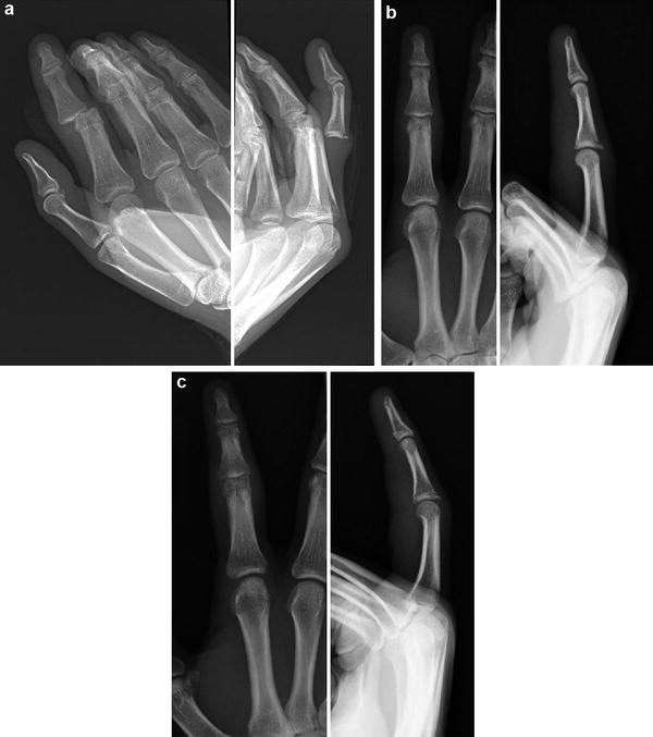
Fig. 6
(a) AP and lateral of 16-year-old male with index PIP fracture dislocation. Note the small epiphyseal fragment dorsally. (b) 16-year-old male 2 weeks status postreduction of the index PIP joint. (c) 16-year-old male 4 weeks status postreduction of the index PIP dislocation. Note that the dorsal epiphyseal fragment has healed
Classification
Dislocations are classified based on the degree and direction of displacement. It is standard to refer to the more distal bone in describing the dislocation. For example, a dislocation of the middle phalanx dorsal to the proximal phalanx is a dorsal dislocation. Dislocations should be thought of as complete or incomplete and simple or complex. In incomplete injuries the base of the phalanx remains perched on the head of the adjacent phalanx. In complete injuries the phalanges are bayoneted, as the base has dislocated completely and is located next to the head of the adjacent phalanx. Incomplete and complete injuries have also been described as type I and type II, though this is a less useful classification and is not commonly used. In this scheme, a type III injury would be a fracture dislocation. Simple versus complex dislocations describe whether the dislocation is reducible by closed versus open means, respectively. Avulsion fractures may be considered under the Salter-Harris classification and are Salter-Harris type III injuries. Incomplete and complete injuries have also been described as Type I and Type II (Eaton 1971). Fracture-dislocations have been referred to as type III. This numerical system is less commonly used.
Outcome Tools
Outcomes after interphalangeal dislocation are generally scored by range of motion, as a flexion contracture is the most common complication. To date, there are no specific outcome scores to assess finger dislocations.
Treatment Options for Dorsal Dislocation of the Proximal Interphalangeal Joint
Nonoperative Management
Indications/Contraindications
Nonoperative management is generally preferred for these injuries. Patients may require operative treatment for unsuccessful closed reduction attempts. Chronic injuries may also require open reduction with release of scarred or contracted tissues. Fracture dislocations may require operative treatment, depending on the fracture present (Table 7).
Table 7
Proximal interphalangeal joint dislocations | |
|---|---|
Nonoperative treatment | |
Indications | Contraindications |
Successful closed reduction | Closed reduction unsuccessful |
Chronic dislocation (trial of closed reduction should be attempted)
Stay updated, free articles. Join our Telegram channel
Full access? Get Clinical Tree
 Get Clinical Tree app for offline access

| |