Chapter 561 Goiter
A goiter is an enlargement of the thyroid gland. Persons with enlarged thyroids can have normal function of the gland (euthyroidism), thyroid deficiency (hypothyroidism), or overproduction of the hormones (hyperthyroidism). Goiter may be congenital or acquired, endemic, or sporadic.
The goiter often results from increased pituitary secretion of thyroid-stimulating hormone (TSH) in response to decreased circulating levels of thyroid hormones. Thyroid enlargement can also result from infiltrative processes that may be inflammatory or neoplastic. Goiter in patients with Graves disease and thyrotoxicosis is caused by thyrotropin receptor–stimulating antibodies (TRSAbs).
561.1 Congenital Goiter
Congenital goiter is usually sporadic and can result from a fetal thyroxine (T4) synthetic defect or from administration of antithyroid drugs or iodides during pregnancy for the treatment of maternal thyrotoxicosis. Goitrogenic drugs and iodides cross the placenta and at high doses can interfere with synthesis of thyroid hormone, resulting in goiter and hypothyroidism in the fetus. The concomitant administration of thyroid hormone with the goitrogen does not prevent this effect, because insufficient amounts of T4 cross the placenta. Iodides are included in many proprietary cough preparations used to treat asthma; these preparations should be avoided during pregnancy because they have often been reported to cause congenital goiter. Amiodarone, an antiarrhythmic drug with 37% iodine content, has also caused congenital goiter with hypothyroidism. Even when the infant is clinically euthyroid, there may be retardation of osseous maturation, low levels of T4, and elevated levels of TSH. In women with Graves disease receiving antithyroid drugs, these effects can occur when the mother takes propylthiouracil at only 100-200 mg/24 hr; all such infants should undergo thyroid studies at birth. Administration of thyroid hormone to affected infants may be indicated to treat clinical hypothyroidism, to hasten the disappearance of the goiter, and to prevent brain damage. Because the condition is rarely permanent, thyroid hormone may be safely discontinued after the antithyroid drug has been excreted by the neonate, usually after 1-2 wk.
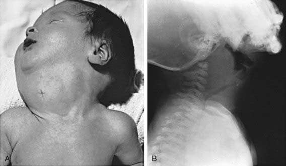
Enlargement of the thyroid at birth may occasionally be sufficient to cause respiratory distress that interferes with nursing and can even cause death. The head may be maintained in extreme hyperextension. When respiratory obstruction is severe, partial thyroidectomy rather than tracheostomy is indicated (Fig. 561-1).

Figure 561-1 Congenital goiter in infancy. A, Large congenital goiter in an infant born to a mother with thyrotoxicosis who had been treated with iodides and methimazole during pregnancy. B, A different infant, 6 wk old, with increasing respiratory distress and cervical mass since birth. The operation revealed a large goiter that almost completely encircled the trachea. Notice the anterior deviation and posterior compression of the trachea. Partial thyroidectomy completely relieved the symptoms. It is apparent why a tracheostomy is not adequate treatment for these infants. The cause for the goiter was not found.
Goiter is almost always present in the infant with neonatal Graves’ disease. These goiters usually are not large; the infant manifests clinical symptoms of hyperthyroidism. The mother often has a history of Graves disease, although discovery of neonatal hyperthyroidism can lead to the diagnosis of maternal Graves’ disease. Thyroid enlargement results from transplacental passage of maternal thyroid-stimulating immunoglobulin (Chapter 562.1). TSH receptor-activating mutations are also a recognized cause of congenital goiter and hyperthyroidism.
When no causative factor is identifiable, a defect in synthesis of thyroid hormone should be suspected. Neonatal screening programs find congenital hypothyroidism caused by such a defect in 1/30,000-50,000 live births. It is advisable to treat immediately with thyroid hormone and to postpone more-detailed studies for later in life. If a specific defect is suspected, genetic tests to identify a mutation may be undertaken (Chapter 559). Because these defects are transmitted by recessive genes, a precise diagnosis is helpful for genetic counseling. Monitoring subsequent pregnancies with ultrasonography can be useful in detecting fetal goiters (Chapter 90).
Iodine deficiency as a cause of congenital goiter is rare in developed countries but persists in isolated endemic areas (see later). More important is the recognition that severe iodine deficiency early in pregnancy can cause neurologic damage during fetal development, even in the absence of goiter. The iodine deficiency can result in maternal and fetal hypothyroidism, preventing the partially protective transfer of maternal thyroid hormones.
When the “goiter” is lobulated, asymmetric, firm, or large to an unusual degree, a teratoma within or in the vicinity of the thyroid must be considered in the differential diagnosis (Chapter 563).
Bizhanova A, Kopp P. Minireview: the sodium-iodide symporter NIS and pendrin in iodide homeostasis of the thyroid. Endocrinol. 2009;150:1084-1090.
Caron P, Moya CM, Malet D, et al. Compound heterozygous mutations in the thyroglobulin gene (1143delC and 6725G → A [R2223H]) resulting in fetal goitrous hypothyroidism. J Clin Endocrinol Metab. 2003;88:3546-3553.
Hashimoto H, Hashimoto K, Suehara N. Successful in utero treatment of fetal goitrous hypothyroidism: case report and review of the literature. Fetal Diagn Ther. 2006;21:360-365.
Koksal N, Akturk B, Saglan H, et al. Reference values for neonatal thyroid volumes in a moderately iodine-deficient area. J Endocrinol Invest. 2008;31:642-646.
Moreno JC, Klootwijk W, van Toor H, et al. Mutations in the iodotyrosine deiodinase gene and hypothyroidism. N Engl J Med. 2008;358:1811-1818.
Remuzzi G, Garattni S. Elimination of iodine-deficiency disorders in Tibet. Lancet. 2008;371:1980-1981.
Teng W, Shan Z, Teng X, et al. Effect of iodine intake on thyroid diseases in China. N Engl J Med. 2006;354:2782-2893.
Zimmernamm MB, Jooste PL, Pandav CS. Iodine-deficiency disorders. Lancet. 2008;372:1251-1262.
561.2 Intratracheal Goiter
One of the many ectopic locations of thyroid tissue is within the trachea. The intraluminal thyroid lies beneath the tracheal mucosa and is often continuous with the normally situated extratracheal thyroid. The thyroid tissue is susceptible to goitrous enlargement, which involves the normally situated and the ectopic thyroid. When there is obstruction of the airway associated with a goiter, it must be ascertained whether the obstruction is extratracheal or endotracheal. If obstructive manifestations are mild, administration of sodium L-thyroxine usually causes the goiter to decrease in size. When symptoms are severe, surgical removal of the endotracheal goiter is indicated.
561.3 Endemic Goiter and Cretinism
Etiology
The association between dietary deficiency of iodine and the prevalence of goiter or cretinism is well established. A moderate deficiency of iodine can be overcome by increased efficiency in the synthesis of thyroid hormone. Iodine liberated in the tissues is returned rapidly to the gland, which resynthesizes triiodothyronine (T3) preferentially at a higher rate than normal. This increased activity is achieved by compensatory hypertrophy and hyperplasia (goiter), which satisfy the demands of the tissues for thyroid hormone. In geographic areas where deficiency of iodine is severe, decompensation and hypothyroidism can result. It is estimated that 1 billion persons in developing countries live in areas of iodine deficiency.

Stay updated, free articles. Join our Telegram channel

Full access? Get Clinical Tree

