18.2 Glomerulonephritis, renal failure and hypertension
Haematuria
Isolated microscopic haematuria
Asymptomatic isolated microscopic haematuria is common and can be detected on a single occasion in 0.5–2% of school-aged children. The majority of these children will not have significant kidney disease and, in the absence of symptoms or other abnormalities on urinalysis, further investigation is required only if haematuria is documented in at least three different specimens taken over a period of 2–3 weeks.
Persistent microscopic haematuria is usually benign, provided there is no infection or proteinuria, renal function is normal and no structural abnormality is present on ultrasonography. Possible diagnoses include idiopathic hypercalciuria, thin membrane disease or a mild proliferative glomerulonephritis such as immunoglobulin A (IgA) nephropathy (see below). A family history of microscopic haematuria is suggestive of thin basement membrane disease, which is often inherited in an autosomal dominant fashion.
Glomerulonephritis
The clinical features of glomerulonephritis are:
• acute fluid overload – oedema, pulmonary oedema, congestive cardiac failure
• renal impairment – oliguria, raised plasma creatinine level.
Acute presentation with these clinical features is seen most commonly in post-streptococcal glomerulonephritis. Other forms of glomerulonephritis in childhood may have a less severe onset (Box 18.2.1). Most forms of glomerulonephritis result from an immunologically mediated injury involving either deposition of circulating immune complexes in the glomerulus or a specific antibody to the glomerular basement membrane.
Post-streptococcal glomerulonephritis
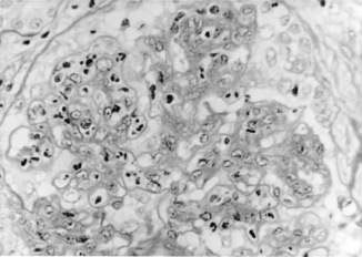
This disorder follows 7–14 days after group A β-haemolytic streptococcal throat infection and 3–6 weeks after streptococcal skin infection. It is hypothesized that streptococcal antigens deposit in glomeruli with activation of the complement system. The pathological appearance consists of proliferation of mesangial and endothelial cells with neutrophil infiltration (Fig. 18.2.1). Crescents may be present. Immunofluorescence shows IgG and C3, and electron-dense deposits (humps) are demonstrated by electron microscopy.
![image](/wp-content/uploads/2016/08/B9780702042928000591_f18-03-9780702042928.jpg)
Fig. 18.2.1 Mesangial proliferation and neutrophil infiltration in post-streptococcal glomerulonephritis (periodic acid–Schiff stain, original magnification ×800).
Clinically this disorder usually presents with macroscopic haematuria, acute fluid overload and hypertension in a school-aged child. Lassitude, fever and loin pain also may be present. Physical examination may reveal hypertension, papilloedema, facial and leg oedema. Serum urea, creatinine and potassium concentrations are often raised, and urinalysis shows red blood cell casts and dysmorphic red cells. Mild normocytic normochromic anaemia is common and is due to haemodilution from fluid overload. The anti-streptolysin O titre (ASOT) and anti-streptococcal DNase B are raised in 90% of cases. Activation of the classical complement pathway leads to low serum levels of C3, which generally returns to normal within 6–12 weeks.
The major complications of acute post-streptococcal glomerulonephritis are secondary to acute kidney injury resulting in salt and water retention. Fluid overload is responsible for hypertension and in severe cases can result in hypertensive encephalopathy or left ventricular failure. Fluid overload is best managed initially with furosemide 2–4 mg/kg daily, fluid restriction and a low-salt diet. More severe hypertension may be treated with oral nifedipine or prazosin. Bed rest is necessary only when the blood pressure is increased. A course of oral penicillin for 10 days eradicates any existing streptococcal infection, but does not alter the natural history of this condition.
The period of oliguria lasts for up to 10 days and dialysis is indicated in cases where the blood urea rises above 50–60 mmol/L, or when hyperkalaemia or pulmonary oedema is not controlled by diuretics and fluid restriction. The long-term prognosis is excellent, with only 1% developing chronic kidney disease. Microscopic haematuria may continue for up to 2 years, but proteinuria should clear within 6 months. Renal biopsy is not indicated unless there is uncertainty of diagnosis with the initial investigations or the period of oliguria lasts for longer than 3 weeks.
Other infectious agents including viral and bacterial organisms rarely can produce an illness similar to post-streptococcal nephritis. These organisms include staphylococci and Pneumococcus, and Echo, Coxsackie and Epstein–Barr viruses.
IgA nephropathy
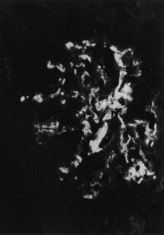
This glomerulopathy is present in 50% of children who have recurrent episodes of macroscopic haematuria. The episodes of haematuria often occur simultaneously with intercurrent viral infections and may be associated with flank pain. Other presentations include abnormal urinalysis on medical examination and, rarely, an acute glomerulonephritis with renal failure. Renal biopsy shows a focal proliferative glomerulonephritis with IgA in the mesangium (Fig. 18.2.2). Although the prognosis for most children with IgA nephropathy is good, long-term studies show that about 10% progress to chronic renal failure by 15 years after onset of disease. Bad prognostic features include impaired renal function at presentation, heavy proteinuria and hypertension.
Henoch–Schönlein purpura
This disease is a vasculitic illness involving predominantly small vessels in the skin, large joints and gastrointestinal tract (see Chapter 16.2). The illness is preceded by upper respiratory tract infection in 30–50% of patients. These children present with a petechial or purpuric rash that is localized over lower limbs and buttocks, abdominal pain and arthritis. A mild nephritis with microscopic haematuria and proteinuria is seen in 50–70% of cases. Rarely, blood pressure and serum creatinine levels are raised. Renal histology shows a proliferative glomerulonephritis with IgA in the mesangium. The prognosis is good, with less than 5% developing chronic kidney disease.
Lupus erythematosus
Systemic lupus erythematosus (SLE) presents in childhood in 20% of cases and is more common in children from Asian countries. Facial rash, arthritis and fever are common presenting symptoms. Serum C3 complement is usually low and anti-nuclear antibodies can often be detected, especially to double-stranded DNA. Renal biopsy is indicated if haematuria and proteinuria are present. The type of glomerular disease in SLE can vary from a mild focal proliferative glomerulonephritis to a diffuse crescentic glomerulonephritis. The treatment is complex but generally involves various combinations of immunosuppressant medications such as prednisolone, azathioprine, cyclophosphamide or mycophenolate. The amount of immunosuppression is dependent on clinical severity of kidney disease and renal histology. This disorder is discussed further in Chapter 13.3.
Alport syndrome
This is a familial disorder in production of type IV collagen and is inherited as an X-linked dominant (85% of cases), autosomal dominant or autosomal recessive condition. In males, this disorder presents in the first 10 years of life with microscopic or macroscopic haematuria and proteinuria, and is followed by onset of renal failure in the teenage years. High-tone nerve deafness and eye abnormalities are the other features of the syndrome.
Proteinuria
Isolated proteinuria
Transient proteinuria can be seen in many conditions including fever, exercise and seizures, and disappears when the condition resolves. Proteinuria that is detected in the standing position but not when recumbent is known as orthostatic or postural proteinuria; this occurs in 10% of children and is more common in adolescence. Testing with urinary dipsticks or urine protein : creatinine ratio shows negligible amounts of protein in first daytime void and increased protein excretion during the day. This phenomenon is benign but proteinuria in an overnight urine specimen will usually require biopsy to determine the cause.
Nephrotic syndrome
Nephrotic syndrome is defined as:
• proteinuria (> 40 mg per m2 per h or protein/creatinine ratio > 250 mg/mmol)
• hypoalbuminaemia (< 25 mg/dL)
The annual incidence in children is approximately 2–4 per 100 000. The major conditions associated with a primary nephrotic syndrome are listed in Box 18.2.2.
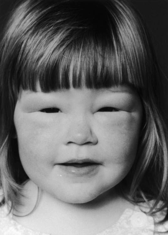
Minimal change nephrotic syndrome
The majority of children present between the ages of 1 and 4 years with generalized oedema (Fig. 18.2.3). Renal biopsy is not initially indicated if clinical features suggest minimal change disease (Box 18.2.3).
![](https://freepngimg.com/download/social_media/63059-media-icons-telegram-twitter-blog-computer-social.png)
Stay updated, free articles. Join our Telegram channel
![](https://clinicalpub.com/wp-content/uploads/2023/09/256.png)
Full access? Get Clinical Tree
![](https://videdental.com/wp-content/uploads/2023/09/appstore.png)
![](https://videdental.com/wp-content/uploads/2023/09/google-play.png)