Chapter 505 Glomerulonephritis Associated with Infections
505.1 Acute Poststreptococcal Glomerulonephritis
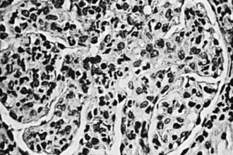
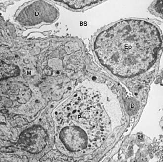
Group A β-hemolytic streptococcal (GAS) infections are common in children and can lead to the postinfectious complication of acute glomerulonephritis (GN). Acute poststreptococcal glomerulonephritis (APSGN) is a classic example of the acute nephritic syndrome characterized by the sudden onset of gross hematuria, edema, hypertension, and renal insufficiency. It is one of the most common glomerular causes of gross hematuria in children and is a major cause of morbidity in GAS infections.
Etiology and Epidemiology
APSGN follows infection of the throat or skin by certain “nephritogenic” strains of GAS. Epidemics and clusters of household cases occur throughout the world, and 97% of cases occur in less-developed countries. The overall incidence has decreased in industrialized nations, presumably due to improved hygienic conditions. Poststreptococcal GN commonly follows streptococcal pharyngitis during cold-weather months and streptococcal skin infections or pyoderma during warm-weather months. Although epidemics of nephritis have been described in association with throat (serotype 12) and skin (serotype 49) infections, this disease is most commonly sporadic.
Pathology
The kidneys appear symmetrically enlarged. Glomeruli appear enlarged and relatively bloodless and show diffuse mesangial cell proliferation, with an increase in mesangial matrix (Fig. 505-1). Polymorphonuclear leukocyte infiltration is common in glomeruli during the early stage of the disease. Crescents and interstitial inflammation may be seen in severe cases, but these changes are not specific for poststreptococcal GN. Immunofluorescence microscopy reveals a pattern of “lumpy-bumpy” deposits of immunoglobulin and complement on the glomerular basement membrane (GBM) and in the mesangium. On electron microscopy, electron-dense deposits, or “humps,” are observed on the epithelial side of the GBM (Fig. 505-2).

Figure 505-1 Glomerulus from a patient with poststreptococcal glomerulonephritis appears enlarged and relatively bloodless and shows mesangial proliferation and exudation of neutrophils (×400).

Figure 505-2 Electron micrograph in poststreptococcal glomerulonephritis demonstrating electron-dense deposits (D) on the epithelial cell (Ep) side of the glomerular basement membrane. A polymorphonuclear leukocyte (P) is present within the lumen (L) of the capillary. BS, Bowman space; M, mesangium.
Pathogenesis
Morphologic studies and a depression in the serum complement (C3) level provide strong evidence that ASPGN is mediated by immune complexes. Precise mechanisms by which nephritogenic streptococci induce immunologic injury continue to be elucidated. Several mechanisms of immune injury have been supported by experimental models and human studies and include circulating immune complex formation with streptococcal antigens and subsequent glomerular deposition, molecular mimicry whereby circulating antibodies elicited by streptococcal antigens react with normal glomerular antigens, in situ immune complex formation of antistreptococcal antibodies with glomerular deposited antigen, and complement activation by directly deposited streptococcal antigens.
Group A streptococci possess M proteins, and nephritogenic strains are related to the M protein serotype. The search for the precise nephritogenic antigen suggests that streptococcal pyogenic exotoxin (SPE B) and nephritis-associated streptococcal plasmin receptor (NAPlr) are promising candidates. Both have been identified in glomeruli of affected patients, and in 1 study, circulating antibodies to SPE B were found in all patients. Cross-reactivity of SPE B and other M proteins with various components of the glomerular basement membrane also give evidence for molecular mimicry.
Clinical Manifestations
Poststreptococcal GN is most common in children aged 5-12 yr and uncommon before the age of 3 yr. The typical patient develops an acute nephritic syndrome 1-2 wk after an antecedent streptococcal pharyngitis or 3-6 wk after a streptococcal pyoderma. The history of a specific infection may be absent, because symptoms may have been mild or have resolved without patients receiving specific treatment or seeking the care of a medical provider.

Stay updated, free articles. Join our Telegram channel

Full access? Get Clinical Tree

