Fig. 1
Modified Mallet Score (Courtesy of Shriners Hospital for Children, Philadelphia)
Additional assessment of shoulder and upper extremity function can take into account patient and family reports of function. The Pediatric Outcomes Data Collection Instrument (PODCI) is a tool that measures function and quality of life in several domains and has been validated in the BPBP population (Dedini et al. 2008). The use of this measure can help to guide long-term functional goals and track improvements over time.
Imaging and Other Diagnostic Studies
The complex three-dimensional anatomy of the shoulder girdle, combined with the largely cartilaginous nature of the glenohumeral joint in young children, limits the utility of plain radiographs in the assessment of the shoulder joint following BPBP. The most important information to be gained from imaging the shoulder includes the shape of the glenoid and the position of the humeral head relative to the glenoid. The progression of glenohumeral dysplasia has been well described (Waters et al. 1998) and tends to progress from increased glenoid retroversion to complete posterior dislocation of the humeral head and severe dysplasia of the glenoid. Pseudoglenoid formation and humeral head flattening further complicate the dysplasia. These deformities are most pronounced in the axial plane, requiring imaging in this plane.
Ultrasonography has been widely used for assessment of hip dysplasia in the infant and can be easily applied to assessment of glenohumeral dysplasia in the same age group (Moukoko et al. 2004). Using a posterior axial approach with a linear array transducer, the humeral head and posterior glenoid can be routinely visualized in children up to 15–18 months of age. The relative position of the humeral head and glenoid can be assessed, allowing assessment of glenohumeral subluxation and dislocation. Ultrasonography has two distinct advantages over MRI. First, it does not require sedation or anesthesia, as is typically required for MRI in the infant. Second, it can be a dynamic study, assessing the relative position of the humeral head and glenoid throughout a range of glenohumeral motion. However, the use of ultrasound to assess the shape of the glenoid has not yet been proven reliable. Nonetheless, ultrasonography can be used as a screening tool to detect infantile glenohumeral dislocation.
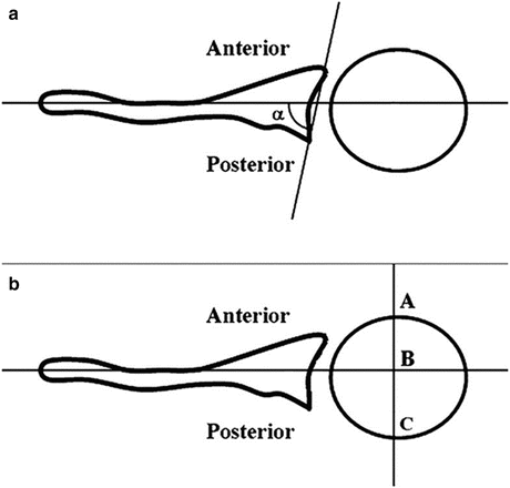
MRI provides the most comprehensive evaluation of the immature glenohumeral joint following BPBP. The cartilaginous glenoid, humeral head, and labrum can be visualized, allowing quantitative measurement of glenoid retroversion and percent posterior subluxation of the humeral head (Fig. 2) (Waters et al. 1998). In addition, the muscles of the shoulder girdle can be visualized, allowing at least a qualitative evaluation of muscle atrophy. The disadvantage of this imaging modality is the need for sedation or general anesthesia to allow a motionless study in the young child. Computed tomography (CT) can be utilized in the older child with an ossified humeral head and glenoid, often obviating the need for sedation or anesthesia, although ionizing radiation is used.


Fig. 2
(a) Glenoid retroversion measurements (Glenoid version = [alpha]-90°) and (b) humeral head subluxation measurements (PHHA = AB/AC × 100 %) (From Lippert et al. (2012))
Nonoperative Management
Nonoperative treatment in the form of occupational and physical therapy remains the cornerstone of initial management of residual shoulder dysfunction and serves as the only treatment required for a majority of children. Goals of therapy involve maintenance of passive joint range motion while awaiting neurological recovery, together with functional rehabilitation following neurological recovery and assistance with motor skill development.
As passive external rotation in adduction correlates with glenohumeral dysplasia, maintenance of this motion is especially important in therapy. The brachium is stabilized against the chest wall, and the shoulder is passively externally rotated using the flexed elbow as a lever arm. Exercises are performed several times with each diaper change, using basic principles of passive stretching. Additional passive motion in the glenohumeral joint is important but difficult to maintain. The abduction contracture is particularly challenging to stretch given the difficulty of adequately stabilizing the scapula. Joint mobilization techniques can be used to maintain a supple glenohumeral joint despite scapular hypermobility, but such techniques can be difficult for parents and caregivers to perform at home.
Aside from maintaining passive range of motion, motor reeducation is vitally important in therapy. Because of the initial weakness of the deltoid and supraspinatus, abduction attempts recruit scapulothoracic movement. Scapular taping may assist with reacquisition of glenohumeral motion by partially inhibiting compensatory scapulothoracic movement. Similarly, the trumpet sign (abduction of the shoulder during hand-to-mouth movement) may initially be due to weakness of external rotation and elbow flexion but may become habitual despite improved muscle strength. Therefore, therapy is necessary to optimize the pattern of movement, not just the strength of individual muscles.
Botulinum toxin A injection has become an increasingly accepted adjunct to physical and occupational therapy in other pediatric neurological disorders, and it may have a role in BPBP. Brachial plexus injury, as a peripheral nerve injury, does not result in muscle spasticity. However, botulinum toxin has been used to facilitate motor learning in recovering muscles following BPBP by temporarily weakening antagonists. A series of eight children underwent botulinum toxin injections into the triceps, pectoralis major, and/or latissimus dorsi muscles, with immediate and 4-month improvements in function of the opposing reinnervating muscles (DeMatteo et al. 2006). Similarly, four patients with biceps-triceps co-contraction demonstrated improved active elbow flexion 18 months following a single botulinum toxin injection into the triceps muscle (Heise et al. 2005). In addition, botulinum toxin injection in the internal rotators may ease the passive stretching of external rotation in adduction by weakening the resistance and discomfort associated with these stretching exercises in the young child. Such a strategy has been used in infants with posteriorly subluxated shoulders, allowing successful reduction that was maintained at 1 year in 24 of 35 patients in one series (Ezaki et al. 2010). The indications for botulinum toxin injection in the setting of BPBP require further elucidation.
Operative Treatment
Surgical treatment of the secondary shoulder dysfunction following BPBP aims to accomplish three goals: (1) restoration of passive motion by contracture release, (2) realignment of the dysplastic glenohumeral joint, and (3) augmentation of muscle power in the weak domains of shoulder movement. If these goals cannot be accomplished, palliative surgery in the form of humeral osteotomy can improve global shoulder function without addressing the glenohumeral joint deformity and dysfunction.
A variety of procedures have been described for surgical release of the shoulder internal rotation contracture and augmentation of external rotation strength. These procedures, modified over many decades, typically involve lengthening or sectioning internal rotators, such as the subscapularis and/or pectoralis major, and transfer of functioning muscles, such as the latissimus dorsi and/or teres major to the posterior or posterosuperior rotator cuff, to augment external rotation and abduction function. Many earlier series have demonstrated short- and medium-term gains in global shoulder function but with results that tend to deteriorate over time, potentially due to a historical lack of awareness of glenohumeral dysplasia.
The increasing awareness of glenohumeral dysplasia over the past decade has created an opportunity to evaluate the effects of these surgical procedures on glenohumeral dysplasia progression. In 2005, Waters and Bae reported a series of 25 children who underwent latissimus dorsi and teres major tendon transfers with or without lengthening of the subscapularis or pectoralis major (Waters and Bae 2005). At 2-year follow-up, a five-point improvement in global shoulder function on the Mallet scale was noted, in keeping with previous reports, but only modest improvements were seen in glenoid retroversion and glenohumeral subluxation on MRI or CT imaging. The authors concluded that muscle rebalancing surgery only halted the progression of dysplasia but did not allow substantial remodeling. Similarly, Kozin et al. in 2006 described a complete lack of glenohumeral remodeling at 1-year follow-up in 23 children treated with a similar combination of procedures (Kozin et al. 2006). However, that same year, a series of 33 children was reported describing 2-year follow-up of arthroscopic release of the subscapularis with or without latissimus dorsi transfer (Pearl et al. 2006). Of 15 children for whom preoperative and follow-up MRI scans were available, 12 showed substantial glenohumeral remodeling. Also published in 2006 was a retrospective study of 109 patients who underwent open subscapularis release and teres major transfer (El-Gammal et al. 2006). Follow-up CT scans available at least 1 year postoperatively in 39 patients demonstrated glenoid retroversion that correlated positively with age at surgery, with normal glenoid retroversion in children operated prior to 4 years of age and no glenohumeral subluxation in children operated by 2 years of age. These two reports, in contrast to the prior two, suggested that remodeling may indeed be possible.
Validation of remodeling potential has now been provided by a number of recent reports. Waters and Bae recently reported a series of 23 patients who underwent subscapularis/pectoralis major lengthenings and latissimus dorsi/teres major transfers but with the addition of open glenohumeral reduction (Waters and Bae 2009). In these patients, 83 % demonstrated glenohumeral remodeling at 2 years, with significant improvements in both glenoid retroversion and glenohumeral subluxation. Similarly, Kozin et al. adopted the technique of arthroscopic subscapularis release with or without external rotation tendon transfers and reported a series of 44 children with significant remodeling on 1-year follow-up MRI (Kozin et al. 2010). These reports draw attention to strategic adaptations in surgical technique that place increased importance on obtaining appropriate glenohumeral articular alignment in addition to extra-articular muscle balance.
Contracture Release/Reduction
The treatment of the shoulder internal rotation contracture following BPBP has been modified over the years (Fairbank 1913; Sever 1916; L’Episcopo 1934; Hoffer et al. 1978; Pearl and Edgerton 1998; Waters and Bae 2005; Newman et al. 2006; Pearl et al. 2006; Pedowitz et al. 2007). Contemporary techniques for treatment of the shoulder internal rotation contracture include (1) open or arthroscopic sectioning or lengthening of the subscapularis and/or pectoralis major tendons with or without release of the anterior glenohumeral joint capsule. The relative advantages and disadvantages of open versus arthroscopic release have been debated, but no clear winner has emerged. Similarly, no clear indications exist to select one technique over the other, as each technique has been reported to successfully improve passive external rotation. The true differences may be borne out with longer follow-up evaluation of glenohumeral alignment and remodeling, as some techniques of contracture release may allow better articular realignment than others. Nonetheless, surgical treatment of the internal rotation contracture should be considered when (1) the internal rotation contracture progresses to less than 20° of passive external rotation in adduction despite appropriate nonoperative means and/or (2) glenohumeral dislocation or progressive glenohumeral dysplasia is documented by axial imaging. Internal rotation contracture release should be used with caution in patients who have substantial functional deficits in midline function, as many reports have demonstrated a risk of worsening internal rotation function following internal rotation contracture release (van der Sluijs et al. 2004; Kambhampati et al. 2006; Kozin et al. 2006; Newman et al. 2006; Pearl et al. 2006).
Techniques
Subscapularis Slide
Surgical release of the subscapularis from its origin on the anterior surface of the scapula has been used for decades, with its proponents citing a low rate of loss of internal rotation function, since the insertion of the subscapularis is left intact (Carlioz and Brahimi 1971). However, opponents cite a high rate of contracture recurrence. The scapula is approached through a longitudinal incision along the lateral border of the scapula beginning superiorly from the posterior axillary fold. The lateral inferior angle of the scapula is retracted laterally from the wound, and the subscapularis is elevated extraperiosteally from its anterior surface beginning inferiorly and progressing superiorly. Adequate release requires elevation of the superior muscle belly deep in the wound, and this portion is performed by palpating tight bands as the shoulder is passively externally rotated. The release is considered sufficient when the shoulder has 60–80° of passive external rotation in adduction. The wound is closed with absorbable sutures, and the arm is positioned in a shoulder spica cast in external rotation and 20–30° of abduction. This procedure can also be accomplished through a longitudinal incision at the medial border of the scapula, releasing the subscapularis beginning medially and progressing laterally.
Arthroscopic Partial Subscapularis Tenotomy
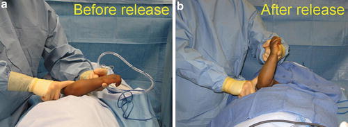
The patient is positioned in the lateral decubitus position. Care must be taken during positioning to protect the contralateral brachial plexus with an axillary roll and to protect all bony prominences with adequate padding. The shoulder is examined under anesthesia to confirm the preoperative internal rotation contracture. In the case of an infantile dislocation, the shoulder is imaged ultrasonographically under anesthesia using the posterior axial view described by Moukoko et al. (2004). The arthroscope (1.9 mm for infants, 2.7 mm for older children) is inserted through a standard posterior portal. If the joint is dislocated, the portal entry is located more medially than typical, with a more laterally directed trajectory of the arthroscope. Care must be taken to avoid injury to the soft, unossified humeral head, and familiarity with shoulder arthroscopy is essential. Following joint inspection and identification of the intra-articular subscapularis tendon, an up-biting basket resector is inserted through an anterior portal, and the superior 2–3 mm of subscapularis tendon is sectioned. The shoulder is then passively externally rotated in adduction to spread the cut fibers, sliding them along the intact fibers (Fig. 3). The release is considered successful if at least 60° of passive external rotation in adduction can be achieved (Fig. 4). If necessary, additional fibers of the subscapularis are sectioned until the desired passive external rotation is achieved, but neither the joint capsule nor the entire intra-articular subscapularis tendon is sectioned. In the case of an infantile dislocation, the shoulder is again imaged with a sterilely draped ultrasound probe to confirm glenohumeral reduction in external rotation. Following successful release, the tendons of the latissimus and teres major muscles are transferred – if indicated – through a transverse axillary approach to the posterosuperior rotator cuff, as described below (Waters 2005).


Fig. 3
Surgical technique of arthroscopic partial subscapularis release, as viewed from the posterior portal in a left shoulder

Fig. 4
Intraoperative photographs demonstrating maximum passive external rotation in adduction before (a) and after (b) arthroscopic partial subscapularis release
Postoperatively, the shoulder is immobilized in a shoulder spica cast in adduction and external rotation following a release alone or in external rotation and abduction following a release and tendon transfers. The cast is removed at 4 weeks postoperatively, and occupational therapy is begun. No brace is used following cast removal, unless the contracture begins to recur.
Open Subscapularis Release
The subscapularis can be lengthened or partially released at its insertion through open approaches as well. An anterior deltopectoral approach grants access to the extra-articular portion of the subscapularis tendon and has been used for z-lengthenings of this tendon (van der Sluijs et al. 2004). However, residual weakness of internal rotation commonly complicated such a lengthening procedure, and it has fallen out of favor. The subscapularis can also be approached through a transverse axillary incision used for the latissimus and teres major tendon transfer as described below. Once the latissimus tendon is harvested from the humeral shaft for transfer, the joint capsule can be seen in the deep portion of the wound. An anteroposterior incision in the inferior capsule allows direct inspection of the glenohumeral joint. The intra-articular, superior portion of the subscapularis tendon can be released with or without the anterior joint capsule. The glenohumeral joint can be anteriorly translated for reduction of a posterior dislocation under direct visualization. As this approach is typically used simultaneously with muscle transfers, the shoulder spica cast applied postoperatively is typically placed with the arm in abduction and external rotation.
Release of Other Structures
Other structures have been implicated in contracture pathophysiology, and several authors have advocated for release of these structures as well. The pectoralis major tendon may be tight, restricting passive external rotation in abduction, and this tendon can be z-lengthened through an anterior deltopectoral approach or axillary approach as described above. Additionally, the corocohumeral ligament and coracoid have been found by some to be impediments to reduction of the dislocated and dysplastic glenohumeral joint, and these structures can be released or removed through an anterior approach. In a long-standing dislocation, the anterior joint capsule may require sectioning, and others have advocated reefing of the posterior joint capsule as well (Waters and Bae 2009). However, unlike adhesive capsulitis in the adult shoulder, the joint capsule is not routinely viewed as the primary structure responsible for the contracture.
Abduction Contracture Release
Only very recently have surgeons begun to address the glenohumeral abduction contracture that has been recognized for decades (Putti 1932). Several techniques have been reported, including partial deltoid release, subscapularis origin release, and acromion osteotomy, but published clinical studies are lacking.
Shoulder internal rotation contracture release |
|---|
Preoperative planning |
OR table: Standard table or beach chair if preferred for arthroscopy |
Position/positioning aids: Lateral decubitus in bean bag with axillary roll for subscapularis slide; lateral decubitus or beach chair for arthroscopic release, based on preference |
Fluoroscopy location: No fluoroscopy, but portable ultrasonography unit placed in front of patient to allow visualization from a posterior transverse approach to confirm reduction in the setting of an infantile glenohumeral dislocation |
Equipment: Standard soft tissue surgical equipment. Small joint 1.9 mm or 2.7 mm arthroscope, small joint instruments |
Tourniquet (sterile/nonsterile): N/A |
Subscapularis slide for shoulder internal rotation contracture |
|---|
Surgical steps |
A longitudinal incision is made along the lateral border of the scapula beginning superiorly from the posterior axillary fold |
The lateral inferior angle of the scapula is retracted laterally from the wound, and the subscapularis is elevated extraperiosteally from its anterior surface beginning inferiorly and progressing superiorly |
Adequate release requires elevation of the superior muscle belly deep in the wound, and this portion is performed by palpating tight bands as the shoulder is passively externally rotated |
The release is considered sufficient when the shoulder has 60–80° of passive external rotation in adduction |
The wound is closed with absorbable sutures |
Arthroscopic partial subscapularis tenotomy for shoulder internal rotation contracture |
|---|
Surgical steps |
The arthroscope (1.9 mm for infants, 2.7 mm for older children) is inserted through a standard posterior portal. If the joint is dislocated, the portal entry is located more medially than typical, with a more laterally directed trajectory of the arthroscope |
Following joint inspection and identification of the intra-articular subscapularis tendon, an up-biting basket resector is inserted through an anterior portal and the superior 2–3 mm of subscapularis tendon is sectioned |
The shoulder is then passively externally rotated in adduction to spread the cut fibers, sliding them along the intact fibers |
The release is considered successful if at least 60° of passive external rotation in adduction can be achieved |
If necessary, additional fibers of the subscapularis are sectioned until the desired passive external rotation is achieved, but neither the joint capsule nor the entire intra-articular subscapularis tendon is sectioned |
In the case of an infantile dislocation, the shoulder is again imaged with a sterilely draped ultrasound probe to confirm glenohumeral reduction in external rotation |
The portals are closed with Steri-Strips or absorbable sutures |
Shoulder internal rotation contracture release |
|---|
Postoperative protocol |
Type of immobilization: Shoulder spica cast in 30–40° of abduction and 40–60° of external rotation |
Length of immobilization: 4 weeks |
Rehab protocol: Preoperative therapy is restarted after removal of cast with emphasis on passive external rotation in adduction. Consider external rotation splint a night for early recurrence of contracture |
Muscle Transfers
Persistent weakness of abduction and external rotation is very common following incompletely resolved BPBP. If nerve surgery (nerve grafting, nerve transfers) has not been performed or has not restored adequate power in the C5 distribution, secondary muscle transfers can reliably improve global shoulder function in abduction and external rotation. Such procedures have been described for decades and have evolved over time. The basic principles remain the same, however. An abundance of adductors and internal rotators that remain innervated provides an opportunity to restore balance about the glenohumeral joint by transferring one or more of these functioning muscles to the posterior humerus or rotator cuff to allow them to function as external rotators. The most commonly transferred muscles are the latissimus dorsi and teres major. The relative improvement in abduction versus external rotation can be selected by the location to which the muscles are transferred. If improved external rotation in adduction is desired, the tendons can be inserted on the posterior humeral shaft, as described by L’Episcopo (1934). If the goal is to improve abduction as well as external rotation in abduction (hand-to-neck on the Mallet scale), then the tendons can be transferred to the posterosuperior rotator cuff as described by Hoffer and Phipps (1998). Therefore, the exact technique of the operation can be varied based on the indication. It is important to note that improving external rotation in adduction should not sacrifice functional internal rotation in adduction. Loss of midline function from internal rotation weakness is more functionally disabling than a lack of external rotation in adduction. Therefore, a contraindication to external rotation tendon transfer is a preexisting inability to internally rotate to the sagittal midline, as such a surgery would worsen that function further.
Stay updated, free articles. Join our Telegram channel

Full access? Get Clinical Tree


