Essentials of Diagnosis
- • Scrotal pain and swelling is typically unilateral in epididymitis.
- • Epididymitis is typically characterized by the presence of urethritis or bacteriuria.
- • An abnormally high position of the testicle may indicate testicular torsion.
- • Have a low threshold to obtain a doppler ultrasound or radionuclide scanning to rule out testicular torsion in adolescents or young adults, because prompt surgical intervention is essential to save the involved testicle.
General Considerations
Epididymitis, an inflammatory process involving the epididymis, is one of the primary etiologies of the acute scrotum syndrome. Epididymitis is common and can cause substantial short-term morbidity (eg, suffering and loss of time from work) and long-term complications (eg, infertility, chronic epididymitis, etc). The incidence of epididymitis may range from 1 to 4 per 1000 men per year. The inflammatory process causes a gradual onset of scrotal pain and swelling that is characteristically unilateral.
With the improved understanding of the etiology of epididymitis, the diagnosis and management of this condition is becoming more rational, leading to decreased morbidity and, possibly, to prevention of recurrences. Epididymitis usually results from infection. There are two main infectious causes: (1) urethral infection with Neisseria gonorrhoeae or Chlamydia trachomatis, and (2) genitourinary tract infection with coliform bacteria or Pseudomonas aeruginosa (see Table 6–1). Age is an important predictor of the etiology, with heterosexual men younger than 35 years of age more likely to have a sexually transmitted pathogen, and older individuals more likely to have a pathogen associated with bacteriuria. In rare cases, epididymitis may occur as a complication of systemic infection with various bacterial, fungal, viral, or parasitic pathogens, or may be due to noninfectious causes (see Table 6–1). In prepubertal boys, epididymitis may be related to concomitant presence of structural, functional, or neurologic abnormalities of the genitourinary tract. Men who have sex with men and who practice insertive anal intercourse are at greater risk for epididymitis caused by coliform bacteria. Cases for which no etiologic agent can be determined after thorough investigation are referred to as idiopathic.
| Associated with urethritis |
| Gonorrhea |
| Chlamydia |
| Trichomoniasis |
| Associated with bacteriuria |
| Coliform bacteria (eg, Escherichia coli) |
| Pseudomonas aeruginosa |
| Associated with funguria |
| Candida spp |
| Associated with systemic infection |
| Bacterial |
| Tuberculosis |
| Mycobacterium other than M tuberculosis (MOTT) |
| Brucellosis |
| Haemophilus influenzae |
| Fungal |
| Histoplasmosis |
| Coccidioidomycosis |
| Blastomycosis |
| Cryptococcosis |
| Viral |
| Mumps |
| Cytomegalovirus |
| Parasitic |
| Schistosomiasis |
| Sparganosis |
| Bancroftian filariasis |
| Associated with drugs |
| Amiodarone |
| Associated with systemic vasculitis |
| Behçet syndrome |
| Henoch-Schönlein purpura |
| Polyarteritis nodosa |
| Wegener granulomatosis |
| Associated with postinfectious etiology |
| Upper respiratory tract infections |
| Associated with trauma |
Pathogenesis
The epididymis is a sausage-shaped structure positioned on the posterior aspect of the testicle. It consists of a single, delicate convoluted tubule 12–15 feet long. During passage through the epididymis, sperm become motile and achieve the potential to fertilize an ovum. Hence, inflammation and fibrosis from epididymitis can impair the passage and maturation of sperm, leading to infertility. In epididymitis associated with urethritis or bacteriuria, there is a retrograde spread of infection intraluminally to the epididymis. In contrast, systemic infections spread to the epididymis by a hematogenous route. Reflux of sterile urine does not cause epididymitis.
Clinical Findings
Clinical manifestations, laboratory results, and imaging findings may differ, depending on the etiology of the acute scrotum syndrome. In epididymitis, clinical findings are influenced by whether the etiology is infectious versus noninfectious, and in the case of the former, by whether the presentation is local versus systemic.
Patients with epididymitis characteristically complain of testicular or scrotal pain and may also complain of inguinal pain. In severe cases, acute swelling of the spermatic cord may result in flank pain from obstruction of the ureter as it crosses over the spermatic cord. More than two thirds of patients with epididymitis describe a gradual onset of pain.
Symptoms typical of the underlying cause, such as urethral discharge associated with sexually transmitted epididymitis or symptoms of urinary urgency and frequency associated with urinary tract infection, are discussed in detail below. Rarely, epididymitis may present with nonspecific symptoms such as fever or malaise, especially in patients with epididymitis associated with chronic catheterization or neurogenic bladder associated with a spinal cord injury.
On examination, the scrotum on the involved side may be red and edematous. The testicle tends to lie in the normal position in the scrotum. Shortly after the onset of inflammation, the tail of the epididymis, which connects with the vas deferens near the lower pole of the testes, is swollen. Later, swelling spreads to the head of the epididymis, near the upper pole of the testes. The groove between the epididymis and the testicle should be examined, as this will help to demonstrate whether the maximum swelling is in the testicle or in the epididymis. The spermatic cord may be swollen and tender (a condition termed funiculitis). A hydrocele may be present; characteristically this results from secretion of fluid by the inflamed tunica vaginalis. Signs and symptoms more specific for a particular etiology of epididymitis are discussed below.
Men with epididymitis secondary to sexually transmitted pathogens often have a history of dysuria or urethral itching or discharge. Usually, they have a history of recent sexual exposure. If the patient has not voided recently, spontaneous urethral discharge may be apparent on examination. It is important to recognize that asymptomatic urethral infection without discharge may occur. Such asymptomatic urethral infections have been estimated for up to 50% of gonococcal infections and over 75% of chlamydial infections. If no spontaneous discharge is noted, then the urethra should be stripped from the base of the penis to the urethral meatus (“milked”) and examined again. In some patients, digital rectal examination may reveal abnormalities suggestive of bacterial prostatitis. The degree of scrotal erythema and epididymal edema may be less in patients with chlamydial epididymitis compared with those in whom epididymitis is a result of other etiologies. However, massive erythema and edema may also occur with untreated C trachomatis epididymitis.
In patients with coliform or pseudomonal epididymitis, a history of bacteriuria or symptoms suggesting urinary tract infection may or may not be present. Symptoms include urinary frequency, urgency, or dysuria. Patients may have a history of symptoms suggestive of urinary tract obstruction (eg, hesitancy or slow urinary stream), indicating conditions that predispose them to urinary tract infection (eg, urethral stricture and benign prostatic hypertrophy). Others may have a history of conditions predisposing them to bacteriuria, including prostatic calculi, recent genitourinary or prostate instrumentation, neurogenic bladder, an indwelling catheter, or chronic bacterial prostatitis.
Bilateral epididymal involvement is more common in epididymitis caused by systemic diseases; in contrast, epididymitis associated with urethritis or bacteriuria is nearly always unilateral. Symptoms or signs relating to systemic infection or inflammation may be present. For example, in tuberculous epididymitis, patients can present with clinical disease involving the kidneys, adrenal glands, lymphatics (retroperitoneal, abdominal, or mediastinal), or all of these structures. Patients with tuberculous epididymitis often have a prior history of pulmonary tuberculosis or a history of exposure to tuberculosis. Another example is epididymitis occurring with Behçet syndrome, in which patients may have oral or genital ulcers, other cutaneous lesions, eye involvement (eg, iritis, uveitis, etc), arthritis, and central nervous system involvement. Genitourinary examination findings in systemic infectious or inflammatory disorders otherwise tend to resemble the findings in epididymitis associated with urethritis or bacteriuria. One exception is that in tuberculous epididymitis, a “string of beads” may be noted on palpation of the vas deferens due to presence of granulomas. An additional, less specific, finding is prostatic calculi that may be detected on digital rectal examination.
Sterile epididymitis has been recognized as a complication of amiodarone therapy, especially at high dosages. Although amiodarone-associated epididymitis is usually bilateral, it may be unilateral.
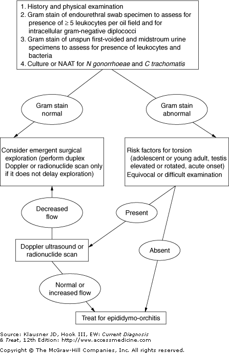
Because the vast majority of cases of epididymitis result from urethritis or bacteriuria, laboratory evaluation should target these entities, unless the history or clinical findings suggest a different entity. Recommended evaluations are discussed below and summarized in Figure 6–1.
Either urethral Gram stain or urine microscopy, and preferably both of these evaluations, are indicated routinely in the initial evaluation of suspected epididymitis (see Figure 6–1).
In cases of suspected urethritis, Gram-stained smear of an endourethral swab specimen will often indicate the presence of urethritis (≥5 polymorphonuclear leukocytes per oil field). The Gram stain can also establish, with a high degree of certainty, whether the etiology is gonococcal (ie, demonstrating gram-negative diplococci) or nongonococcal.
Ideally, first-voided and midstream urine specimens should be examined for bacteria and white blood cells. Comparison of the urinary sediments in the first-voided and midstream specimens may reveal the source of pyuria (ie, urethra or bladder); higher concentrations of leukocytes are typically found in the first-voided urine of patients with urethritis.
A midstream urine Gram stain should be performed, which can be used to presumptively establish the diagnosis of bacteriuria. Presence of more than 1 gram-negative rod per high-power field of unspun midstream urine correlates with presence of more than 105 organisms per milliliter.
If microscopy is not available, then dipstick urinalysis can be performed on a first-voided urine specimen. A positive leukocyte esterase test is consistent with an etiology of urethritis or bacteriuria but does not distinguish between these two possibilities.
All urethral specimens should be tested for N gonorrhoeae and C trachomatis. Culture has been the traditional diagnostic standard for chlamydia and gonorrhea; however, tests of first-voided urine for N gonorrhoeae and C trachomatis using highly sensitive and specific nucleic acid amplification tests may prove just as or even more useful, although some experts believe more experience with these tests is needed in men with epididymitis.
Quantitative midstream urine culture should be obtained in all cases of acute epididymitis in which bacteriuria is suspected. Culture identifies the responsible pathogen and also provides antimicrobial susceptibility data.
Finding so-called sterile pyuria in individuals with negative gonococcal and chlamydial tests who have not recently received antimicrobial therapy should prompt the clinician to consider alternative infectious or inflammatory etiologies, such as mycobacterial, other bacterial, or fungal epididymitis. Pyuria is absent in drug-associated epididymitis and in most cases of epididymitis associated with autoimmune disorders.
Imaging is not indicated in routine cases of epididymitis. However, in the selected situations considered below, imaging procedures may prove very helpful.


