Chapter 296 Echinococcosis (Echinococcus granulosus and Echinococcus multilocularis)
Etiology
Echinococcosis (hydatid disease or hydatidosis) is the most widespread, serious human cestode infection in the world (Fig. 296-1). Two major Echinococcus species are responsible for distinct clinical presentations, E. granulosus (cystic hydatid disease) and the more malignant E. multilocularis (alveolar hydatid disease). The adult parasite is a small (2-7 mm) tapeworm with only 2-6 segments that inhabits the intestines of dogs, wolves, dingoes, jackals, coyotes, and foxes. These carnivores pass the eggs in their stool, which contaminates the soil, pasture, and water, as well as their own fur. Domestic animals such as sheep, goats, cattle, and camels ingest E. granulosus eggs while grazing. Humans are also infected by consuming food or water contaminated with eggs or by direct contact with infected dogs. The larvae hatch, penetrate the gut, and are carried by the vascular or lymphatic systems to the liver, lungs, and less commonly bones, brain, or heart.

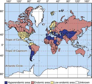
Figure 296-1 Worldwide distribution of cystic echinococcosis.
(From McManus DP, Zhang W, Li J, et al: Echinococcosis, Lancet 362:1295–1304, 2003.)
E. granulosus shows high intraspecific variation. One distinct variant is found in a sylvatic wolf/moose cycle in North America and Siberia. The transmission cycle of E. multilocularis is similar to that of E. granulosus, except that this species is mainly sylvatic and uses small rodents as its natural intermediate hosts. The rodents are consumed by foxes, their natural predators, and sometimes by dogs and cats.
Epidemiology
There is potential for transmission of this parasite to humans wherever there are herd animals and dogs. Even in urban areas, dogs may be infected by eating entrails after home slaughter of domestic animals. Cysts have been detected in up to 10% of the human population in northern Kenya and Western China. In South America, the disease is prevalent in sheepherding areas of the Andes, the beef-herding areas of the Brazilian/Argentine Pampas, and Uruguay. Among developed countries, the disease is recognized in Italy, Greece, Portugal, Spain, and Australia, and is reemergent in dogs in Great Britain. In North America, transmission occurs by way of the sylvatic cycle in Alaska, Canada, and Isle Royale on Lake Superior, as well as in foci of the domestic cycle in sheep raising areas of western USA.
Transmission of E. multilocularis occurs primarily in temperate climates of Northern Europe, Siberia, Turkey, and China. Transmission is decreasing among native peoples in Alaska and Canada as dogs are replaced by mechanized forms of transportation. A separate species, Echinococcus vogeli, causes polycystic disease similar to alveolar hydatidosis in South America.
Pathogenesis
In areas endemic for E. granulosus, the parasite is often acquired in childhood, but liver cysts require many years to become large enough to detect or cause symptoms. In children, the lung is a common site, whereas in adults 70% of cysts develop in the right lobe of the liver. Cysts can also develop in bone, the genitourinary system, bowels, subcutaneous tissues, and brain. The host surrounds the primary cyst with a tough, fibrous capsule. Inside this capsule, the parasite produces a thick lamellar layer with the consistency of a soft-boiled egg white. This layer supports a thin germinal layer of cells responsible for production of thousands of juvenile-stage parasites (protoscolices) that remain attached to the wall or float free in the cyst fluid. Smaller internal daughter cysts may develop within the primary cyst capsule. The fluid in a healthy cyst is colorless, crystal clear, and watery. After medical treatment or with bacterial infection, it may become thick and bile stained.
Infection with E. multilocularis

Stay updated, free articles. Join our Telegram channel

Full access? Get Clinical Tree

