22.1 Ear, nose and throat disorders
Paediatric otolaryngologists see children with mucosal diseases of the upper aerodigestive tract (ears, nose, oral cavity, pharynx and larynx), airway obstruction, and pathologies of communication.
• foreign bodies, in any of these areas
• otitis media, both acute and chronic
• hearing loss, both conductive and sensorineural, and congenital and acquired
• rhinosinusitis and its complications
• tonsillitis and suppuration in the neck
Growth and development
In infants the face is relatively small compared with the cranium, and elongation occurs with mandibular and maxillary growth as the permanent teeth erupt, from about 6 years of age. This coincides with development of the eustachian tube, as it becomes more vertical and functions more efficiently. During this time there is also growth and maturation of the immune system, with enlargement of the tonsils and adenoids; these increase in size until the age of about 7 years, and then start to involute (Fig. 22.1.1).
![image](/wp-content/uploads/2016/08/B9780702042928000712_f22-01-9780702042928.jpg)
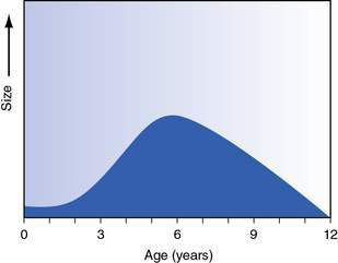
Fig. 22.1.1 Adenoidal size in relation to age.
Redrawn with permission from Dhillon RS, East CA, 2006 Ear, Nose and Throat and Head and Neck Surgery: An Illustrated Colour Text, 3e. With permission from Elsevier.
Infants are obligate nose-breathers until approximately 5 months, when they are able to mouth-breathe. The nose is small with considerable airway resistance and this explains why infants have difficulty breathing and feeding with a cold.
If there is obstruction at the back of the nose with choanal atresia, intervention is needed in the newborn period, especially if the obstruction is bilateral.
The paranasal sinuses also develop with facial growth. The ethmoid and maxillary sinuses are present at birth; the sphenoid sinus is aerated at 5 years and the frontal sinus at 10 years of age. This has important implications for the complications of acute sinusitis: ethmoid sinusitis can develop intraorbital complications from infancy, but intracranial extension from frontal and sphenoid sinusitis is rare under the age of 10 years.
The temporal bone contains the outer, middle and inner ears. The inner ear is adult size at birth. In the newborn the tympanic membrane (TM) is more horizontal and so difficult to see, and it becomes vertical with growth. At birth there is only one mastoid air cell (the antrum), but there is rapid pneumatization after this.
The paediatric larynx is higher in young children than in adults, and the epiglottis may be seen on tongue protrusion. The larynx is cone-shaped, so the subglottis is the narrowest part and therefore prone to damage and stenosis in prolonged intubation. The laryngeal cartilages are soft and tend to collapse in.
The larynx grows rapidly until the age of 3 years, and then slows before another rapid growth at puberty; the vocal cords lengthen and the angle of the cartilages change, and this accounts for the voice changes that occur.
Trauma and foreign bodies
Young children can place, inhale or ingest a variety of foreign bodies into each orifice. Small batteries become moist and erode mucosa and cartilage, so need to be removed urgently.
The ear
A foreign body may be wedged in the canal; if this is vegetable matter, it can expand and cause pain. If not removed easily with a head-light and wax curette, it is best to remove under anaesthesia or sedation to avoid further trauma, especially to the TM.
Trauma from a long or sharp foreign body can perforate a TM, and perforation may also result from a direct hit on the ear. If kept dry, these usually heal spontaneously.
Bleeding from the ear occurs with skull-base fractures, and may contain cerebrospinal fluid (CSF). These should be allowed to heal with minimal intervention, to avoid contaminating the CSF. The child should be examined for nystagmus, hearing loss and facial nerve injury.
An audiogram is performed to identify ossicular and inner ear damage in all cases of trauma and perforation.
The nose
A foreign body in the nose may present as unilateral rhinorrhoea with an offensive odour; this resolves once the foreign body is removed. This may be accomplished with the child sitting upright and supported against a parent’s chest. The nose is sprayed with anaesthetic/decongestant and the foreign body is removed by placing a wax curette behind it and pulling it forwards.
Trauma to the nose is common from falling on to a table or step, or sports injury. There is often swelling and bruising initially, and an assessment for deformity may be needed after this has settled. An assessment of the airway and possible septal haematoma should be performed immediately, as it quickly becomes an abscess and pressure on the cartilage causes necrosis and saddle-nose deformity.
Pharynx and oesophagus
A child with a pharyngeal foreign body such as a fish bone may present with drooling and can point to the site; a bone is often lodged in the tonsil and can be readily seen and removed.
If a foreign body is further down or in the oesophagus the child will have dysphagia and regurgitation of saliva, and will need general anaesthesia for removal.
Trauma from running with a stick, pencil or other toy in the mouth is common. If it involves the soft palate, flexible nasopharyngeal examination is performed to ensure there is no injury in the retropharynx, as there is the possibility of air and infection tracking down to the mediastinum. Small punctures of the soft palate will heal well, but if there is a large flap, or the injury is on the free edge of the soft palate, it is sutured under general anaesthesia.
Larynx and bronchi
A child with an inhaled foreign body in the airway may present in the immediate period with choking, gasping and cyanosis, or may have problems later with wheeze, cough and persistent or recurrent pneumonia. A chest X-ray may show ipsilateral hyperinflation from entrapment of air behind the foreign body, but there is a low threshold for performing bronchoscopy if there is a history of choking and no radiological signs. The bronchoscopy is performed under general anaesthesia using a rigid, ventilating bronchoscope and telescopic forceps.
The ear
Congenital abnormalities
Differences in the shape and size of the pinna are common. Corrective surgery is performed after the age of 5 years, when the pinna is near adult size.
There is an association of pinna deformity and middle ear abnormalities; the hearing is tested and appropriate intervention with hearing aids may be needed. Often the loss is conductive and the child can be fitted with a bone-conduction hearing aid.
Major deformities of microtia (small ear) and anotia (absent ear) are managed in interdisciplinary clinics with plastic surgeons to determine timing for surgery, both to correct deformities and to improve hearing, by either a tympanoplasty or bone-anchored hearing aid.
Otitis media
Otitis media is inflammation or infection of the mucoperiosteal lining of the middle ear, and includes the mastoid ear cells and the eustachian tube (ET). There is a spectrum of disease from mild and reversible to chronic and destructive.
• A child with acute otitis media (AOM) usually presents following a cold with severe pain and fever, and may have otorrhoea. Infants and young children may not localize well and present with fever, irritability and sometimes vomiting.
• Otitis media is common in the first year of life, and 75% of children have had at least one episode by the age of 3 years. Recurrent otitis media is defined as at least three episodes in 6 months or four in 12 months. It is most common in autumn and winter as viral infections cause obstruction of the nose and ET.
• The three common causative organisms are Streptococcus pneumoniae, Haemophilus influenzae (non-typeable) and Moraxella catarrhalis.
• The complications of AOM include TM perforation, facial paralysis, mastoiditis, intracranial spread including meningitis and abscess formation, and sigmoid sinus thrombosis.
• A child with mastoiditis has often had symptoms for days before the ear starts to protrude, with erythema and swelling over the mastoid process. The treatment is surgical drainage and antibiotic therapy.
• With unresolved inflammation and eustachian tube obstruction there may be damage and retraction of the TM, with erosion of the ossicles. Retraction pockets form and accumulate keratinizing stratified squamous epithelium, known as a cholesteatoma. This causes bone erosion and usually presents with intermittent otorrhoea with hearing loss. Cholesteatoma may also be congenital and present as a white mass (‘pearl’) behind an intact TM. This may be an incidental finding.
There are some recognized risk factors for otitis media:
• Race – Australian aboriginal children and some Native Americans (Inuit, Apache and Navajo). There are differences in the eustachian tube and immunological response, but socioeconomic factors are also important.
• Craniofacial abnormalities – including cleft palate and Down syndrome.
There are recognized environmental factors, and families can help control these:
• Reduce contact with people with upper respiratory infections, especially large-group childcare centres.
• Avoid tobacco smoke both during and after pregnancy.
• Breastfeed for at least 6 months, preferably 12 months. If bottle-fed, prop the baby up as milk can reflux into the ear if lying flat, causing inflammation.
• Avoid pacifiers/dummies; this is possible from inadvertent sharing in childcare centres.
• Vaccination with the polyvalent pneumococcal vaccine reduces the incidence of AOM by 8%.
Maisie is 15 months old; she has a fever and is crying. She has purulent rhinorrhoea, a bulging left ear drum and fluid in the right ear. Her management includes pain relief and, as she is less than 2 years old, an antibiotic – usually amoxicillin. If not clinically improved in 2 days she should be reviewed to determine whether the acute infection has resolved; if not, she should be changed to a β-lactamase-stable antibiotic such as amoxicillin–clavulanate. After this acute infection Maisie might have fluid in her middle ears for some weeks, without having infection.
Chronic otitis media with effusion (COME)
This is commonly known as ‘glue ear’. ‘Chronic’ implies duration of at least 3 months. There is persistent fluid in the middle ear, usually after AOM, which is asymptomatic apart from hearing loss. The term middle ear effusion (MEE) designates fluid in the ear, without reference to the aetiology or duration.
• COME can be present for 3 months and still resolve spontaneously.
• Biofilms (blankets of bacteria in a low metabolic state and enclosed by a polymeric matrix) may contribute, and the organisms are similar to those that cause AOM.
• There is no evidence that treatment with decongestants, antihistamines, nasal steroids or alternative medications will improve the resolution of MEE.
• The hearing changes in COME are often mild (10–15 dB worse) but there is a wide range, and the criteria for what is a significant loss are uncertain.
• Long-term studies indicate that for children with normal development there are no sequelae for language development from COME.
Thien Phuoc is 4 years old and this winter he has had a lot of colds and two episodes of AOM. He often ignores his mother or asks her to repeat what she has said, but she says he speaks clearly in Vietnamese. On examination, he has fluid in both ears. As there is concern about his hearing Thien Phuoc should have an audiogram, but if it is near the end of winter this can be delayed until summer as his mother is not concerned about his speech, and the fluid may resolve spontaneously. If he has persistent middle ear effusions in summer with a conductive hearing loss, then discussions about whether to insert tubes should start. It may be difficult for health workers to determine whether there is a speech delay in a child who does not speak English.
![](https://freepngimg.com/download/social_media/63059-media-icons-telegram-twitter-blog-computer-social.png)
Stay updated, free articles. Join our Telegram channel
![](https://clinicalpub.com/wp-content/uploads/2023/09/256.png)
Full access? Get Clinical Tree
![](https://videdental.com/wp-content/uploads/2023/09/appstore.png)
![](https://videdental.com/wp-content/uploads/2023/09/google-play.png)