Duodenal Obstruction
Eva Ilse Rubio, MD
DIFFERENTIAL DIAGNOSIS
Common
Malrotation/Midgut Volvulus
Duodenal Atresia
Duodenal Stenosis/Web
Less Common
Jejunal Atresia
Superior Mesenteric Artery (SMA) Syndrome
Duodenal Hematoma
Gastrointestinal Duplication Cyst
Rare but Important
Bezoar
Lymphoma
Annular Pancreas
ESSENTIAL INFORMATION
Key Differential Diagnosis Issues
Differentiate between proximal (high) or distal (low) obstruction in neonates
High (proximal) obstruction
Malrotation/midgut volvulus
Duodenal atresia or stenosis
Duodenal web
Duodenal hematoma
Annular pancreas
Jejunal atresia (proximal portion)
Extrinsic compression from mass or cyst
Mid-level obstruction
Jejunal atresia
Extrinsic compression from mass or cyst
Low (distal) obstruction
Anorectal malformation/anal atresia
Hirschsprung disease
Meconium plug syndrome (small left colon syndrome)
Ileal atresia
Meconium ileus
Extrinsic compression from mass or cyst
Duodenal obstruction falls into proximal (high) obstruction differential
Conventional radiograph may show distention of stomach, duodenum, or a few proximal small bowel loops
Midgut volvulus must be urgently excluded in patients presenting with bilious emesis
UGI is initial radiographic exam for evaluation of proximal (high) obstruction
Helpful Clues for Common Diagnoses
Malrotation/Midgut Volvulus
Malrotation: Abnormal fixation of duodenum to retroperitoneum; prone to twisting upon itself
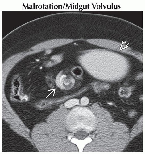
Midgut volvulus: Twisting of malrotated bowel that may result in obstruction, ischemia, and bowel necrosis
Malrotation with midgut volvulus must be excluded in patients who present with bilious emesis
Radiographic findings range from normal abdominal x-ray to distended stomach and proximal duodenum
UGI is next step in work-up
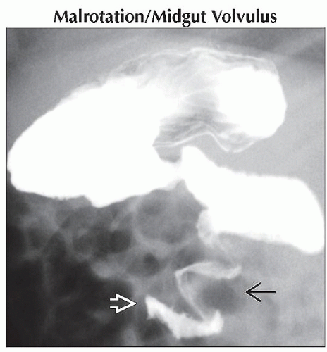
Entire duodenal sweep must course through retroperitoneum on UGI lateral view
Duodenojejunal junction must be situated to left of spinal pedicle and at same level as duodenal bulb
Note: Volvulus may occur in normally rotated patients who have other bowel pathology, such as mass or cyst, adhesions, inspissated meconium, etc.
Duodenal Atresia/Stenosis/Web
Accepted etiology is failure of canalization of lumen in utero rather than vascular insult (as with other atresias)
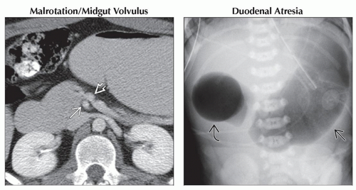
Distended, air-filled stomach and duodenum appear as “double bubble” sign
Distal bowel gas
Usually absent in duodenal atresia
Present in varying amounts in duodenal stenosis or web
Normal caliber colon on contrast enema (unless ileal atresia is present)
Associated with other anomalies
Down syndrome (25%)
Other GI anomalies, such as malrotation, pancreatic and biliary anomalies, Meckel diverticulum, other atresias, gastrointestinal duplications (25%)
Vertebral or rib anomalies (> 33%)
Congenital heart disease
Assess for VACTERL sequence anomalies
Helpful Clues for Less Common Diagnoses
Jejunal Atresia
Few distended loops of bowel may appear as “triple bubble” sign
Intrauterine vascular insult is commonly accepted cause
Associated with other abnormalities
Malrotation/volvulus
Anterior abdominal wall defects
Other, more distal atresias may be present; contrast enema should be performed
If colon caliber is normal, ileal atresia very unlikely
If there is microcolon, other distal (ileal) atresias are present
Superior Mesenteric Artery (SMA) Syndrome
Often in older children with recent history of weight loss
3rd portion of duodenum compressed by overlying superior mesenteric artery
Best diagnosed with upper GI
Proximal half of duodenum often mildly distended
Contrast material has difficulty passing beyond middle of 3rd portion of duodenum, sloshes back and forth
Contrast column halts at vertical linear extrinsic compression caused by SMA
Expect difficulty passing feeding tube beyond this point
Duodenal Hematoma
Typically traumatic in nature
Handlebar injury, direct blow/punch
May result from endoscopy/biopsy
Variable appearance
Focal intramural bulge into lumen
Circumferential wall thickening narrowing lumen
Gastrointestinal Duplication Cyst
Typically round or tubular, highly variable size, commonly present with obstruction
US: Contents may be anechoic, hypoechoic, or complex with debris; may demonstrate bowel wall layers
CT: Low-attenuation contents
Most do not communicate with true bowel lumen
Helpful Clues for Rare Diagnoses
Bezoar
Foreign body bezoar
Appearance is commonly swirled or mottled; depends on what patient has eaten (hair, soil/sand, plant matter, household products)
Lymphoma
Nonspecific soft tissue mass
CT: Homogeneous or mildly heterogeneous, low to intermediate attenuation
US: Homogeneous or mildly heterogeneous, hypoechoic
Annular Pancreas
Usually seen in conjunction with other anomalies, such as duodenal web/stenosis
Image Gallery
 (Left) Axial CECT shows abnormally reversed positions of the mesenteric artery and vein. In this patient with midgut malrotation and volvulus, the mesenteric artery is on the right
 , and the mesenteric vein is on the left , and the mesenteric vein is on the left  . (Right) Frontal radiograph shows a dilated stomach . (Right) Frontal radiograph shows a dilated stomach  and duodenal bulb and duodenal bulb  , together demonstrating the classic “double bubble” sign of duodenal atresia. Note that there is no distal bowel gas. , together demonstrating the classic “double bubble” sign of duodenal atresia. Note that there is no distal bowel gas.Stay updated, free articles. Join our Telegram channel
Full access? Get Clinical Tree
 Get Clinical Tree app for offline access

|


