Chapter 655 Disorders of the Nails
Nail abnormalities in children may be manifestations of generalized skin disease, skin disease localized to the periungual region, systemic disease, drugs, trauma, or localized bacterial and fungal infections (Table 655-1). Nail anomalies are also common in certain congenital disorders (Table 655-2).
Table 655-1 WHITE NAIL OR NAIL BED CHANGES
| DISEASE | CLINICAL APPEARANCE |
|---|---|
| Anemia | Diffuse white |
| Arsenic | Mees lines: transverse white lines |
| Cirrhosis | Terry nails: most of nail, zone of pink at distal end (see Fig. 655-3) |
| Congenital leukonychia (autosomal dominant; variety of patterns) | Syndrome of leukonychia, knuckle pads, deafness; isolated finding; partial white |
| Darier disease | Longitudinal white streaks |
| Half-and-half nail | Proximal white, distal pink azotemia |
| High fevers (some diseases) | Transverse white lines |
| Hypoalbuminemia | Muehrcke lines: stationary paired transverse bands |
| Hypocalcemia | Variable white |
| Malnutrition | Diffuse white |
| Pellagra | Diffuse milky white |
| Punctate leukonychia | Common white spots |
| Tinea and yeast | Variable patterns |
| Thallium toxicity (rat poison) | Variable white |
| Trauma | Repeated manicure: transverse striations |
| Zinc deficiency | Diffuse white |
From Habif TP, editor: Clinical dermatology, ed 4, Philadelphia, 2004, Mosby, p 887.
Table 655-2 CONGENITAL DISEASES WITH NAIL DEFECTS
| Large nails | Pachyonychia congenita, Rubinstein-Taybi syndrome, hemihypertrophy |
| Smallness or absence of nails | Ectodermal dysplasias, nail-patella, dyskeratosis congenita, focal dermal hypoplasia, cartilage-hair hypoplasia, Ellis–van Creveld, Larsen, epidermolysis bullosa, incontinentia pigmenti, Rothmund-Thomson, Turner, popliteal web, trisomy 13, trisomy 18, Apert, Gorlin-Pindborg, long arm 21 deletion, otopalatodigital, fetal alcohol, fetal hydantoin, elfin facies, anonychia, acrodermatitis enteropathica |
| Other | Congenital malalignment of the great toenails, familial dystrophic shedding of the nails |
Abnormalities in Nail Shape or Size
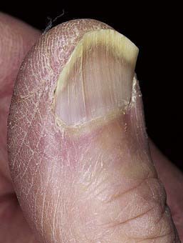
Anonychia is absence of the nail plate, usually a result of a congenital disorder or trauma. It may be an isolated finding or may be associated with malformations of the digits. Koilonychia is flattening and concavity of the nail plate with loss of normal contour, producing a spoon-shaped nail (Fig. 655-1). Koilonychia occurs as an autosomal dominant trait or in association with iron deficiency anemia, Plummer-Vinson syndrome, or hemochromatosis. The nail plate is relatively thin for the first year or two of life and, consequently, may be spoon-shaped in otherwise normal children.

Figure 655-1 Spoon nails (koilonychia). Most cases are a variant of normal.
(From Habif TP, editor: Clinical dermatology, ed 4, Philadelphia, 2004, Mosby, p 885.)
For a discussion of pachyonychia congenita, see Chapter 650.
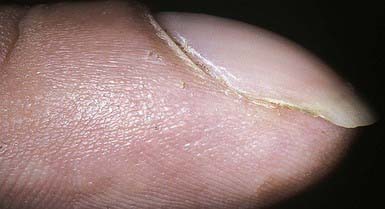
Clubbing of the nails (hippocratic nails) is characterized by swelling of each distal digit, an increase in the angle between the nail plate and the proximal nail fold (Lovibond angle) to >180 degrees, and a spongy feeling when one pushes down and away from the interphalangeal joint, because of an increase in fibrovascular tissue between the matrix and the phalanx (Fig. 655-2). The pathogenesis is not known. Nail clubbing is seen in association with diseases of numerous organ systems, including pulmonary, cardiovascular (cyanotic heart disease), gastrointestinal (celiac disease, inflammatory bowel disease), and hepatic (chronic hepatitis) systems, as well as in healthy individuals as an idiopathic or familial finding.


