Chapter 18 Diseases of the placenta and membranes
ABNORMAL PLACENTATION
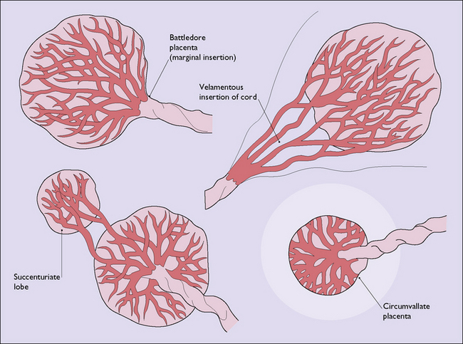
The shape of the definitive placenta is determined at the time the placenta forms, the variations found (Fig. 18.1) mostly having no clinical significance.
The umbilical cord may enter the placenta at its midpoint, may join it at an edge (marginal insertion of the cord), or the umbilical vessels may run some distance along the membranes (velamentous insertion of the cord). Should the vessels run across the cervix they may be compressed by the fetal head during labour, or bleed causing fetal anaemia. In some cases the placenta is smaller than the chorionic plate and the trophoblast invades the decidua laterally more deeply, giving a ridged appearance on the placental surface (placenta circumvallata). In most cases this has no clinical significance, but occasionally may be found in women who have antepartum or intrapartum haemorrhage. In other cases the placenta has an accessory lobe separated by membranes from the main placenta (placenta succenturiata).
In a few cases the trophoblastic invasion is not regulated by maternal immune defences and the myometrium is invaded, causing placenta accreta, increta or percreta. Haemangiomata occur in 1% of placentae. In most cases they are small and of no clinical significance, but larger haemangiomata may be associated with polyhydramnios, antepartum haemorrhage or preterm labour.
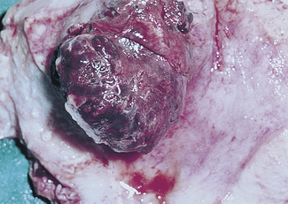
GESTATIONAL TROPHOBLASTIC DISEASE
Gestational trophoblastic disease is uncommon affecting 1/750 pregnancies of women of non-Asian ethnicity but 1/380 of those from Asia. It is potentially lethal, but with treatment a 98% cure rate is attainable. The disease occurs in two forms (Box 18.1).
Hydatidiform mole
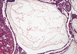
The tumour may have completely or partially replaced the placenta (Fig. 18.2). In the complete form, hydropic swelling and vesicle formation is associated with trophoblastic proliferation and a paucity or absence of blood vessels within the villi (Fig. 18.3). No fetus can be found. Five per cent of complete moles undergo malignant change.

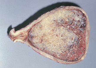
Fig. 18.2 Benign trophoblastic tumour (hydatidiform mole): gross specimen. Note the grape-like masses.

Fig. 18.3 Microphotograph of a benign trophoblastic tumour (hydatidiform mole), showing the distended villi and irregular trophoblastic proliferation.
In the partial form a fetus is present but areas of the placenta show the changes described for the complete mole. Partial moles become malignant less frequently (0.05%).
Malignant trophoblastic disease
The tumour may be confined to the uterus (invasive mole) or spread via the bloodstream to distant organs (choriocarcinoma).
In the invasive mole the trophoblast-covered villi penetrate the myometrial fibres and may extend to other organs, the appearance of the villi remaining that of the benign tumour.
In choriocarcinoma (Fig. 18.4) the tumour is characterized by sheets of trophoblastic cells, both syncytio- and cytotrophoblasts, with few or no villi formed.

Full access? Get Clinical Tree

