Chapter 639 Diseases of the Neonate
Minor evanescent lesions of newborn infants, particularly when florid, may cause undue concern. Most of the entities are relatively common, benign, and transient and do not require therapy.
Sebaceous Hyperplasia
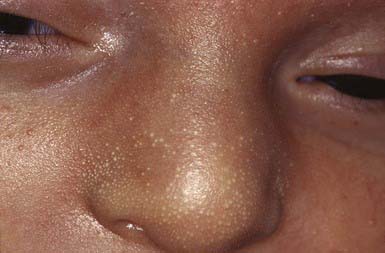
Minute, profuse, yellow-white papules are frequently found on the forehead, nose, upper lip, and cheeks of a term infant; they represent hyperplastic sebaceous glands (Fig. 639-1). These tiny papules diminish gradually in size and disappear entirely within the first few weeks of life.
Milia
Milia are superficial epidermal inclusion cysts that contain laminated keratinized material. The lesion is a firm cyst, 1-2 mm in diameter, and pearly, opalescent white. Milia may occur at any age but in neonates are most frequently scattered over the face and gingivae and on the midline of the palate, where they are called Epstein pearls. Milia exfoliate spontaneously in most infants and may be ignored; those that appear in scars or sites of trauma in older children may be gently unroofed and the contents extracted with a fine-gauge needle.
Sucking Blisters
Solitary or scattered superficial bullae present at birth on the upper limbs of infants at birth are presumably induced by vigorous sucking on the affected part in utero. Common sites are the radial aspect of the forearm, thumb, and index finger. These bullae resolve rapidly without sequelae and should be distinguished from sucking pads (calluses), which are found on the lips in the first few months and are due to combined intracellular edema and hyperkeratosis. The diagnosis can be confirmed by observing the neonate sucking the affected area.
Cutis Marmorata
When a newborn infant is exposed to low environmental temperatures, an evanescent, lacy, reticulated red and/or blue cutaneous vascular pattern appears over most of the body surface. This vascular change represents an accentuated physiologic vasomotor response that disappears with increasing age, although it is sometimes discernible even in older children. Cutis marmorata telangiectatica congenita is clinically similar, but the lesions are more intense, may be segmental, are persistent, and may be associated with loss of dermal tissue, epidermal atrophy, and ulceration.
Harlequin Color Change
A rare but dramatic vascular event, harlequin color change occurs in the immediate newborn period and is most common in low birthweight (LBW) infants. It probably reflects an imbalance in the autonomic vascular regulatory mechanism. When the infant is placed on one side, the body is bisected longitudinally into a pale upper half and a deep red dependent half. The color change lasts only for a few minutes and occasionally affects only a portion of the trunk or face. Changing the infant’s position may reverse the pattern. Muscular activity causes generalized flushing and obliterates the color differential. Repeated episodes may occur but do not indicate permanent autonomic imbalance.
Salmon Patch (Nevus Simplex)
Salmon patches are small, pale pink, ill-defined, vascular macules that occur most commonly on the glabella, eyelids, upper lip, and nuchal area of 30-40% of normal newborn infants. These lesions, which represent localized vascular ectasia,

Stay updated, free articles. Join our Telegram channel

Full access? Get Clinical Tree

