Chapter 649 Diseases of the Epidermis
649.1 Psoriasis
Etiology/Pathogenesis
Psoriasis is characterized by proliferation and abnormal differentiation of keratinocytes and inflammatory cell infiltration of the epidermis and dermis secondary to a primary T-cell abnormality. Psoriasis has a complex multifactorial genetic basis. The major psoriasis-susceptibility gene (PSORS1) is HLA-CW*0602. Numerous other psoriasis susceptibility genes have been identified (PSORS2-PSORS9). Interleukins IL12B and IL23R are the most compelling gene products involved in the pathogenesis of psoriasis.
Clinical Manifestations
This common, chronic skin disorder is first evident in ≈ 30% of affected individuals within the first 2 decades of life. The lesions consist of erythematous papules that coalesce to form plaques with sharply demarcated, irregular borders. If they are unaltered by treatment, a thick silvery or yellow-white scale (resembling mica) develops (Fig. 649-1A). Removal of the scale may result in pinpoint bleeding (Auspitz sign). The Koebner, or isomorphic, response, in which new lesions appear at sites of trauma, is a valuable diagnostic feature. Lesions may occur anywhere, but preferred sites are the scalp, knees, elbows, umbilicus, superior intergluteal fold, and genitals. Small raindrop-like lesions on the face are common. Nail involvement, a valuable diagnostic sign, is characterized by pitting of the nail plate, detachment of the plate (onycholysis), yellowish brown subungual discoloration, and accumulation of subungual debris (Fig. 649-1B).

Figure 649-1 A, Chronic psoriatic plaques. B, Psoriatic nail dystrophy. C, Guttate psoriasis in widespread distribution over the trunk.
Psoriasis is rare in neonates but may be severe and recalcitrant and may pose a diagnostic problem. Other rare forms include psoriatic erythroderma, localized or generalized pustular psoriasis, and linear psoriasis.
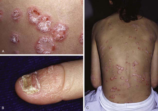
Guttate psoriasis, a variant that occurs predominantly in children, is characterized by an explosive eruption of profuse, small, oval or round lesions that morphologically are identical to the larger plaques of psoriasis (Fig. 649-1C). Sites of predilection are the trunk, face, and proximal portions of the limbs. The onset frequently follows a streptococcal infection; a culture of the throat and serologic titers should be obtained. Guttate psoriasis has also been observed after perianal streptococcal infection, viral infections, sunburn, and withdrawal of systemic corticosteroid therapy.
Differential Diagnosis
Psoriasis is a clinical diagnosis. The differential diagnosis of plaque-type psoriasis includes nummular dermatitis, tinea corporis, seborrheic dermatitis, postinfectious arthritis syndromes, pityriasis rosea, pityriasis lichenoides, and pityriasis rubra pilaris. Scalp lesions may be confused with seborrheic dermatitis, atopic dermatitis, or tinea capitis. Diaper area psoriasis may mimic seborrheic dermatitis, eczematous diaper dermatitis, perianal streptococcal disease, or candidosis. Guttate psoriasis can be confused with viral exanthems, secondary syphilis, pityriasis rosea, pityriasis rubra pilaris, and pityriasis lichenoides chronica (PLC). Nail psoriasis must be differentiated from onychomycosis, lichen planus, and onychodystrophy.
Pathology
When the diagnosis is in doubt, histopathologic examination of an untreated lesion reveals characteristic changes of psoriasis, demonstrating hyperkeratosis, parakeratosis, acanthosis, elongated rete ridges, neutrophilic infiltrate in the epidermis, and lymphocytic infiltrate in the dermis.
Treatment
The therapeutic approach varies with the age of the child, type of psoriasis, sites of involvement, and extent of the disease. Physical and chemical trauma to the skin should be avoided as much as possible (see previous discussion of Koebner response).
The treatment of psoriasis should be viewed as a 4-tier process. The 1st tier is topical therapy. Topical corticosteroid preparations are effective. Mid-potency or stronger topical steroids are necessary (Chapter 638). The preparation that is least potent but effective should be applied twice a day. The topical vitamin D analog calcipotriene is also effective. Calcipotriene can burn and sting, limiting its usefulness in children. One commonly used strategy is to use calcipotriene twice a day on weekdays and a high- to super-potency topical steroid twice a day on weekends. Tazarotene, a topical retinoid, is also useful. It may be used alone or in combination with other topical modalities. Tar preparation and anthralin may also be used. For scalp lesions, applications of a phenol and saline solution (e.g., Baker Cummins P & S Liquid) followed by a tar shampoo are effective in the removal of scales. A high- to super-potency corticosteroid in a foam, solution, lotion, or gel base may be applied when the scaling is diminished. Nail lesions are difficult to treat but may respond to topical tazarotene.
The 3rd tier is systemic therapy. A few children with severe psoriasis require systemic therapy. Methotrexate (0.2 to 0.4 mL/kg once a week), oral retinoids (0.3 to 1.0 mg/kg/day), and cyclosporine (3-5 mg/kg/day) are used for the rare severe and generalized forms of psoriasis. Oral retinoids may be combined with phototherapy.
The 4th tier of therapy is the biologic response modifiers, including the tumor necrosis factor inhibitors etanercept, infliximab, and adalimumab and the T-cell function inhibitors efalizumab and alefacept. Ustekinumab, a human monoclonal antibody that prevents interactions between interleukins IL-12 and IL-23 and their cell surface receptor, has efficacy in treating moderate to severe chronic psoriasis and psoriatic arthritis.
Prognosis
Prognosis is best for children with limited disease. Psoriasis is a lifelong disease characterized by remissions and exacerbations. Arthritis may be an extracutaneous complication.
Anstey A. Home UVB phototherapy for psoriasis. BMJ. 2009;338:1153-1154.
Bartlett BL, Tyring SK. Ustekinumab for chronic plaque psoriasis. Lancet. 2008;371:1639-1640.
Benoit S, Hamm H. Childhood psoriasis. Clin Dermatol. 2007;25:555-562.
Boehncke WH, Boehncke S, Schön MP. Managing comorbid disease in patients with psoriasis. BMJ. 2010;340:200-203.
Burden AD, Boon MH, Leman J, et al. Diagnosis and management of psoriasis and psoriatic arthritis in adults: summary of SIGN guidance. BMJ. 2010;341:c5623.
Cuchacovich RS, Espinoza LR. Ustekinumab for psoriatic arthritis. Lancet. 2009;373:605-606.
Duffin KC, Chandran V, Gladman DD, et al. Genetics of psoriasis and psoriatic arthritis: update and future direction. J Rheumatol. 2008;35:1449-1453.
Griffiths CEM, Strober BE, van de Kerkhof P, et al. Comparison of ustekinumab and etanercept for moderate to severe psoriasis. N Engl J Med. 2010;362(2):118-128.
Kimball AB, Kupper TS. Future perspectives/quo vadis psoriasis treatment? Immunology, pharmacogenomics, and epidemiology. Clin Dermatol. 2008;26:554-561.
Nestle FO, Kaplan DH, Barker J. Psoriasis. N Engl J Med. 2009;361:496-508.
Paller AS, Siegfried EC, Langley RG, et al. Etanercept treatment for children and adolescents with plaque psoriasis. N Engl J Med. 2008;358:241-251.
649.2 Pityriasis Lichenoides
Pityriasis lichenoides encompasses pityriasis lichenoides acuta (PLA; pityriasis lichenoides et varioliformis acuta [PLEVA] and Mucha-Habermann disease) and PLC. The designation of pityriasis lichenoides as acute or chronic refers to the morphologic appearance of the lesions rather than to the duration of the disease. No correlation is found between the type of lesion at the onset of the eruption and the duration of the disease. Many patients have both acute and chronic lesions simultaneously, and transition of lesions from one form into another occurs occasionally. A rare variant, acute febrile ulcernecrotic Mucha-Habermann disease, is also included in the spectrum of pityriasis lichenoides.
Etiology/Pathogenesis
Two main theories exist for the etiology of pityriasis lichenoides. The first is that it arises in a genetically susceptible individual from an atypical immune response to a foreign antigen. The second is that it represents a monoclonal T-cell lymphocytic proliferation on the pathway to cutaneous T-cell dyscrasia.
Clinical Manifestations
Pityriasis lichenoides most commonly manifests in the second and third decades of life; 30% of cases manifest before age 20 yr.
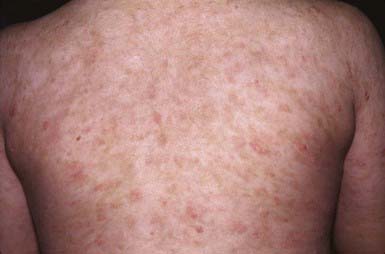
PLC manifests as generalized, multiple, 3- to 5-mm brown-red papules that are covered by a fine grayish scale (Fig. 649-2). Lesions may be asymptomatic or may cause minimal pruritus and occasionally become vesicular, hemorrhagic, crusted, or superinfected. Individual papules become flat and brownish in 2-6 wk, ultimately leaving a hyperpigmented or hypopigmented macule. Scarring is unusual. Lesions are most common on the trunk and extremities and generally spare the face, palmoplantar surfaces, scalp, and mucous membranes.
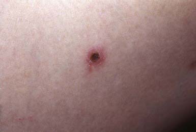
PLA manifests as an abrupt eruption of numerous papules that have a vesiculopustular and then a purpuric center, are covered by a dark adherent crust, and are surrounded by an erythematous halo (Fig. 649-3). Constitutional symptoms such as fever, malaise, headache, and arthralgias may be present for 2-3 days after the initial outbreak. Lesions are distributed diffusely on the trunk and extremities, as in PLC. Individual lesions heal within a few weeks, sometimes leaving a varioliform scar, and successive crops of papules produce the characteristic polymorphous appearance of the eruption, with lesions in various stages of evolution. The overall eruption persists for months to years.
Acute febrile ulceronecrotic Mucha-Habermann disease manifests as fever and ulceronecrotic plaques up to 1 cm in diameter, which are most common on the anterior trunk and flexors of the proximal upper extremities. Arthritis and superinfection of cutaneous lesions with Staphylococcus aureus may also develop. The ulceronecrotic lesions heal with hypopigmented scarring in a few weeks.
Pathology
PLC histologically shows a parakeratotic, thickened corneal layer; epidermal spongiosis; a superficial perivascular infiltrate of macrophages and predominantly CD8 lymphocytes that may extend into the epidermis; and small numbers of extravasated erythrocytes in the papillary dermis.
The histopathologic changes of PLA reflect its more severe nature. Intercellular and intracellular edema in the epidermis may lead to degeneration of keratinocytes. A dense perivascular mononuclear cell infiltrate that extends upward into the epidermis and downward into the reticular dermis, endothelial cell swelling, and extravasation of erythrocytes into the epidermis and dermis are additional characteristic features. The histology of acute febrile ulceronecrotic Mucha-Habermann disease is similar to that of PLA, with leukocytoclastic vasculitis occasionally seen.
Differential Diagnosis
The differential diagnosis of pityriasis lichenoides includes guttate psoriasis, pityriasis rosea, drug eruptions, secondary syphilis, viral exanthems, and lichen planus. The chronicity of pityriasis lichenoides helps preclude pityriasis rosea, viral exanthems, and some drug eruptions. A skin biopsy helps distinguish pityriasis lichenoides from other entities in the differential diagnosis.
Treatment
In general, pityriasis lichenoides should be considered a benign condition that does not alter the health of the child. A lubricant to remove excessive scaling may be all that is necessary if the patient is asymptomatic. Topical steroids may help the pruritus but do not alter the course of the disease. Some children may benefit from treatment with erythromycin (30-50 mg/kg/24hr for 2 mo). Natural sunlight is also helpful. NB-UVB is the treatment of choice for widespread, pruritic disease. The rare febrile ulceronecrotic form may require systemic corticosteroids, other systemic immunosuppressives or anti–tumor necrosis factor or anti–T cell biologic response modifiers.
Khachemoune A, Biyumin ML. Pityriasis lichenoides: pathophysiology, classification, and treatment. Am J Clin Dermatol. 2007;8:29-36.
Sotiriou E, Patsatsi A, Tsorova C, et al. Febrile ulceronecrotic Mucha-Habermann disease: a case report and review of the literature. Acta Derm Venereol. 2008;88:350-355.

Full access? Get Clinical Tree

