Chapter 652 Diseases of Subcutaneous Tissue
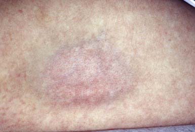
Corticosteroid-Induced Atrophy
Intradermal injection of a corticosteroid can produce deep atrophy accompanied by surface pigmentary changes and telangiectasia (Fig. 652-1). These changes occur approximately 2 wk after injection and may last for months.
652.1 Panniculitis and Erythema Nodosum
Inflammation of fibrofatty subcutaneous tissue may primarily involve the fat lobule or, alternatively, the fibrous septum that compartmentalizes the fatty lobules. Lobular panniculitis that spares the subcutaneous vasculature includes post-steroid panniculitis, lupus erythematosus profundus, pancreatic panniculitis, α1-antitrypsin deficiency, subcutaneous fat necrosis of the newborn, sclerema neonatorum, cold panniculitis, subcutaneous sarcoidosis, and factitial panniculitis. Lobar panniculitis with vasculitis occurs in erythema induratum and, occasionally, as a feature of Crohn disease (Chapter 328.2). Inflammation predominantly within the septum, sparing the vasculature, may be seen in erythema nodosum (Table 652-1 and Fig. 652-2), necrobiosis lipoidica, progressive systemic sclerosis (Chapter 154), and subcutaneous granuloma annulare (Chapter 649). Septal panniculitis that includes inflammation of the vessels is found primarily in leukocytoclastic vasculitis and polyarteritis nodosa (Chapter 161).
Table 652-1 ETIOLOGY OF ERYTHEMA NODOSUM
VIRUSES
Epstein-Barr, hepatitis B, mumps
FUNGI
Coccidioidomycosis, histoplasmosis, blastomycosis, sporotrichosis
BACTERIA AND OTHER INFECTIOUS AGENTS
Group A streptococcus,* tuberculosis,* Yersinia, cat-scratch disease, leprosy, leptospirosis, tularemia, mycoplasma, Whipple disease, lymphogranuloma venereum, psittacosis, brucellosis
OTHER
Sarcoidosis, inflammatory bowel disease,* estrogen-containing oral contraceptives,* systemic lupus erythematosus, Behçet syndrome, severe acne, Hodgkin disease, lymphoma, sulfonamides, bromides, Sweet syndrome, pregnancy, idiopathic*
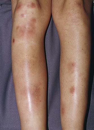
Figure 652-2 Tender red nodules with indistinct borders in a teenage girl with erythema nodosum.
(From Weston AL, Lane AT, Morelli JG: Color textbook of pediatric dermatology, ed 3, St Louis, 2002, Mosby, p 212.)
Erythema Nodosum
Erythema nodosum is a nodular, erythematous hypersensitivity reaction that typically appears with multiple lesions on the exterior surfaces of the arms and legs in the pretibial area (more common) and less often in other cutaneous areas containing subcutaneous fat. The lesions vary in size from 1 to 6 cm, are symmetric, and are oval with the longer axis parallel to the extremity. They initially appear bright or dull red but progress to a brown or purple; they are tense and painful and usually do not ulcerate (see Fig. 652-2). Initial lesions may resolve in 1-2 wk, but new lesions may continue to appear for 2-6 wk. Repeat episodes may occur weeks to months later. Prior to or immediately at the onset of lesions, there may be systemic manifestations that include fever, malaise, arthralgias (50-90%) and rheumatoid factor negative arthritis.
The etiology is unknown in 30-50% of pediatric cases of erythema nodosum; other etiologies are noted in Table 652-1. Group A streptococcal infection and inflammatory disorders (inflammatory bowel disease) are common etiologies in children; sarcoidosis should be considered in young adults.
Post-Steroid Panniculitis
Etiology/Pathogenesis
The mechanism of the inflammatory reaction in the fat in post-steroid panniculitis is unknown.
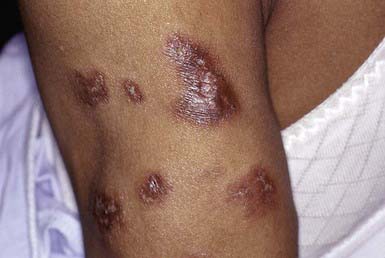
Lupus Erythematosus Profundus (Lupus Erythematosus Panniculitis)
Clinical Manifestations
Lupus erythematosus profundus manifests as one to several firm, well-defined, purple plaques or nodules 1 to 3 cm in diameter, most commonly on the face, buttocks, or proximal extremities. This condition may occur in patients with systemic or discoid lupus erythematosus and may precede or follow the development of other cutaneous lesions. The overlying skin is usually normal but may be erythematous, atrophic, poikilodermatous, or hyperkeratotic (Fig. 652-3). Lesions may be painful and may ulcerate. On healing, a shallow depression generally remains or, rarely, soft pink areas of anetoderma result.
α1-Antitrypsin Deficiency
Etiology/Pathogenesis
Individuals with α1-antitrypsin deficiency have severe homozygous deficiency or, rarely, a partial deficiency of the protease inhibitor α1-antitrypsin, which inhibits trypsin activity and the activity of elastase, serine proteases, collagenase, factor VIII, and kallikrein (Chapter 385). Panniculitis occurs with the Z subtype.


