Development of the Hematopoietic System

Chapter 440 Development of the Hematopoietic System


Hematopoiesis in the Human Embryo and Fetus


Hematopoiesis is the process by which the cellular elements of blood are formed. In the developing human embryo and fetus, hematopoiesis is conceptually divided into 3 anatomic stages: mesoblastic, hepatic, and myeloid. Mesoblastic hematopoiesis occurs in extraembryonic structures, principally in the yolk sac, and begins between the 10th and 14th days of gestation. By 6-8 wk of gestation the liver replaces the yolk sac as the primary site of blood cell production, and during this time the placenta also contributes as a hematopoietic site. By 10-12 wk extraembryonic hematopoiesis has essentially ceased. Hepatic hematopoiesis occurs through the remainder of gestation, although hepatic production diminishes during the 2nd trimester while bone marrow (myeloid) hematopoiesis increases. The liver remains the predominant erythropoietic organ (few if any neutrophils are produced in the human fetal liver) through 20-24 wk of gestation.


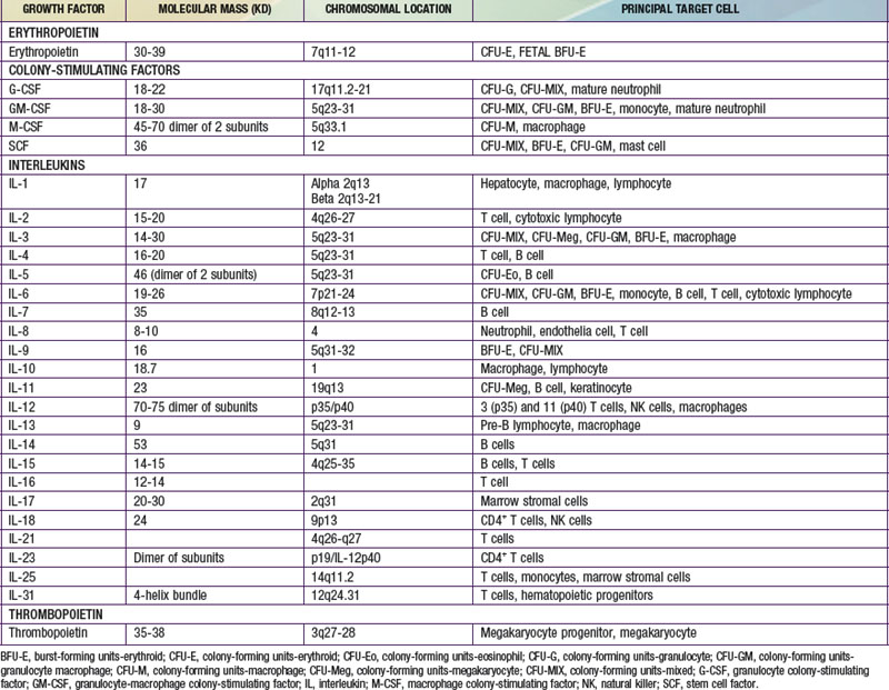
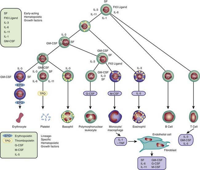
Each hematopoietic organ houses distinct populations of cells. At 18-20 wk, the fetal liver is predominantly an erythropoietic organ, and the marrow produces both erythrocytes and neutrophils. The types of leukocytes present in the fetal liver and marrow differ with gestation. Macrophages precede neutrophils in the marrow, and the ratio of macrophages to neutrophils decreases as gestation progresses. Regardless of gestational age or anatomic location, production of all hematopoietic tissues begins with pluripotent stem cells capable of both self-renewal and clonal maturation into all blood cell lineages. Progenitor cells differentiate under the influence of hematopoietic growth factors (Table 440-1). Fetal hematopoietic growth factor production is independent of maternal growth factor production (Fig. 440-1).


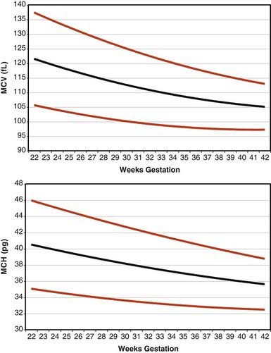
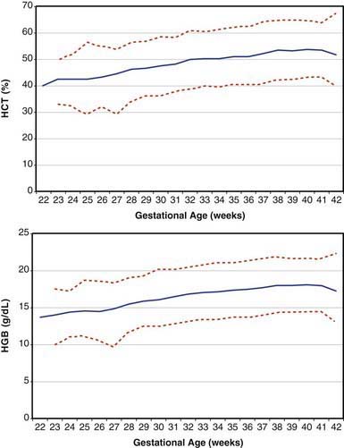
Erythrocytes in the fetus are larger than in adults, and at 22-23 wk gestation the mean corpuscular volume can be as high as 135 fL (Fig. 440-2, upper panel). Similarly the mean corpuscular hemoglobin is very high at 22-23 wk and falls relatively linearly with advancing gestation (see Fig. 440-2, lower panel). In contrast, the mean corpuscular hemoglobin concentration is constant throughout gestation at 34 ± 1 g/fL. Although the size and quantity of hemoglobin in erythrocytes diminish during gestation, the hematocrit and blood hemoglobin concentration gradually increase (Fig. 440-3).


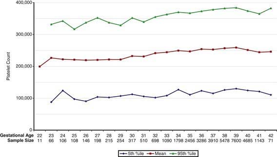
Concentrations of platelets in the blood increase gradually between 22 and 40 wk gestation (Fig. 440-4), but the platelet size, assessed by mean platelet volume, remains constant at 8 ± 1 fL. No differences are observed between males and females in fetal and neonatal reference ranges for erythrocyte indices, hematocrit, hemoglobin, platelet counts, or mean platelet volume measurements.


Fetal Granulocytopoiesis


Neutrophils are first observed in the human fetus about 5 wk after conception as small clusters of cells around the aorta. The fetal bone marrow space begins to develop around the 8th wk after conception, and from 8-10 wk the marrow space enlarges but no neutrophils appear there until 10.5 wk. From 14 wk through term the most common granulocytic cell type in the fetal bone marrow space is the neutrophil. Neutrophils and macrophages originate from a common progenitor cell, but macrophages appear before neutrophils in the fetus, first in the yolk sac, liver, lung, and brain, all before the bone marrow cavity is formed.


Granulocyte colony-stimulating factor (G-CSF) and macrophage colony-stimulating factor (M-CSF) are expressed in developing fetal bone as early as 6 wk post conception, and both are expressed in the fetal liver as early as 8 wk. Granulocyte-macrophage colony-stimulating factor (GM-CSF) and stem cell factor (SCF) also are distributed widely in human fetal tissues. However, no changes in expression of any of these factors, or of their specific receptors, appear to be the signal for fetal production of neutrophils or macrophages, because those signals have not yet been identified.


Fetal blood contains few neutrophils until the 3rd trimester. At 20 wk of gestation the blood neutrophil count is 0-500/mm3. Although mature neutrophils are scarce, progenitor cells with the capacity to generate neutrophil clones are abundant in fetal blood. When cultured in vitro in the presence of recombinant G-CSF, they mature into large colonies of neutrophils. The physiologic role of G-CSF includes upregulating neutrophil production, and this function is present in the fetus and neonate as well as adults. Thus, the low quantities of circulating neutrophils in the 2nd-trimester human fetus may be due, in part, to low production of G-CSF. Monocytes isolated from the blood of adults produce G-CSF when stimulated with a variety of inflammatory mediators such as bacterial lipopolysaccharide (LPS) or interleukin-1 (IL-1). In contrast, monocytes isolated from the blood or organs of fetuses up to 24 wk of gestation generate only small quantities of G-CSF protein and mRNA after LPS or IL-1 stimulation. Despite the low quantities of G-CSF, G-CSF receptors on the surface of neutrophils of newborn infants are equal in number and affinity to those on adult neutrophils.

Stay updated, free articles. Join our Telegram channel

Jun 18, 2016 | Posted by in PEDIATRICS | Comments Off on Development of the Hematopoietic System

Full access? Get Clinical Tree

Get Clinical Tree app for offline access