Fig. 14.1
Dermoid age presentation
Embryology
Acquired implantation. Traumatic implantation of skin into the deeper layers resulting in formation of a dermal cyst lined with squamous epithelium and resembling epidermoid cysts.
Congenital teratomas. Technically from all three types of germinal epithelium (ectoderm, endoderm, and mesoderm), these dermoids, which are usually found in the ovaries or testes, are often included as a subset of teratoma.
Congenital inclusion dermoid cysts. Form along the lines of embryologic fusion and contain both epidermal and dermal elements. These are the lesions found in the head and neck .
Histopathology
Dermoid cysts are benign neoplasms that are derived from epidermal and dermal elements.
Dermoids are usually cystic lesions that contain a cheesy, keratinous substance. They can also be solid tumors, composed of a thick fibrofatty matrix.
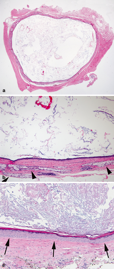
The walls of dermoid cysts are made up of keratinized stratified squamous epithelium along with appendages such as sweat glands sebaceous glands, hair follicles, and connective tissue elements (Fig. 14.2a, b).

Fig. 14.2
Dermoid cyst. a Simple cyst lined by keratinizing squamous epithelium and containing keratin debris and fragments of hair. b Characteristically, the cyst wall contains dermal appendages with well-formed pilosebaceous units (arrowheads). c For comparison, an epidermal inclusion cyst is shown. The lining consists of keratinizing squamous epithelium (arrows) devoid of dermal appendages and the lumen (top) contains laminated keratin debris
The term “dermoid cyst” can be confusing since over the years it has been used to identify several different histopathologic entities. In the head and neck, this term has been applied to (1) true dermoid cysts, (2) epidermal (epidermoid) cysts, and (3) teratoid cysts [5].
Unlike dermoids, the wall of epidermoids contains solely squamous epithelium (Fig. 14.2c). Both of these cyst types usually contain sebaceous debris within their lumen.
Clinically, dermoid and epidermoid cysts behave identically. Differentiation between dermoid and epidermoid cysts can be made only by pathologic examination.
True dermoid cysts are rare. Epidermoid cysts are much more common but because of their similar appearance and location, clinicians often lump the two types of lesions together and call them dermoids.
Teratoid cysts have in the past been confused with dermoids and epidermoids. However, teratoid cysts contain endodermal and/or mesodermal components in addition to their ectodermal-derived epithelium [6].
Presentation
In the most comprehensive study on dermoids to date, New and Erich reviewed 1,495 dermoids cysts treated at the Mayo Clinic from 1910 to 1935 . They noted the majority of dermoid cysts presented in the anal region (44.5 %). This was followed in frequency by the ovaries (42 %) and the head and neck (7 %) [3].
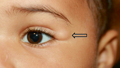
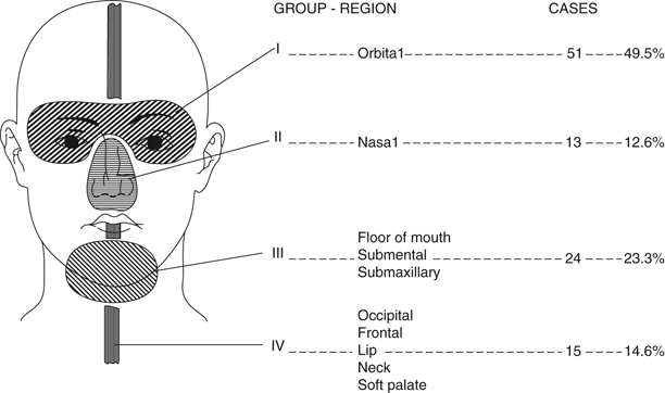
Of the 103 head and neck dermoids reported by New and Erich, the majority were located in the orbital region (Figs. 14.3, 14.4) [3].

Fig. 14.3
Dermoid cyst of lateral canthus region. (Courtesy of Linda Dagi, MD)

Fig. 14.4
Location of head and neck dermoid cysts [3]. (Reprinted with permission from the Journal of the American College of Surgeons, formerly Surgery, Gynecology and Obstetrics)
Stay updated, free articles. Join our Telegram channel

Full access? Get Clinical Tree


