Cystic Kidney
Paula J. Woodward, MD
DIFFERENTIAL DIAGNOSIS
Common
Multicystic Dysplastic Kidney (MCDK)
Hydronephrosis (Mimic)
Obstructive Cystic Dysplasia
Duplicated Collecting System with Obstruction (Mimic)
Rare but Important
Simple Cyst
Meckel-Gruber Syndrome
ESSENTIAL INFORMATION
Key Differential Diagnosis Issues
Do the “cysts” connect?
Real-time evaluation is essential for differentiating an obstructed system from true renal cystic disease
Helpful Clues for Common Diagnoses
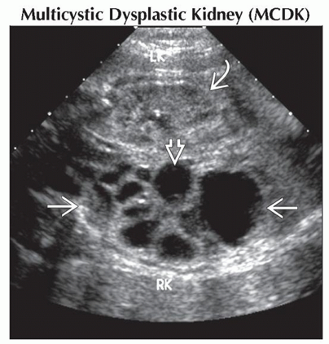
Multicystic Dysplastic Kidney (MCDK)
Multiple, variable-sized cysts in renal fossa
Reniform shape is lost
Often large, distorting normal abdominal anatomy
Variable in-utero course; some involute and others increase in size
Hydronephrosis (Mimic)
Distended calyces may appear “cyst-like”
Must show that calyces connect with renal pelvis (longitudinal views best)
Causes: Ureteropelvic junction (UPJ) or bladder outlet obstruction
Obstructive Cystic Dysplasia
Cystic parenchymal change from chronic obstruction
Hydronephrosis → cortical cysts
Reflects nephron damage and decreased renal function
Cysts are often cortical
Form in subcapsular nephrogenic zone
Kidneys become echogenic
Reniform shape generally retained
Rarely can appear identical to MCDK
Duplicated Collecting System with Obstruction (Mimic)
Upper and lower pole moieties separated by band of renal parenchyma
Upper pole prone to obstruction and can appear as cystic mass
Look for ureterocele in bladder
Helpful Clues for Rare Diagnoses
Simple Cyst
Isolated, unilocular renal cyst
Vast majority resolve during pregnancy
4% progress to MCDK
Meckel-Gruber Syndrome
Triad of classic findings (2 findings required for diagnosis)
Renal cystic dysplasia most consistent finding, present in 95-100%
Grossly enlarged, echogenic kidneys most common, but may present with bilateral, large cysts
Encephalocele in 60-80%
Post-axial polydactyly in 55-75%
Image Gallery
 Coronal ultrasound shows a MCDK on the right
 and a normal kidney on the left and a normal kidney on the left  . A central cyst . A central cyst  could be confused for a renal pelvis, but none of the cysts connected during real-time evaluation. could be confused for a renal pelvis, but none of the cysts connected during real-time evaluation.Stay updated, free articles. Join our Telegram channel
Full access? Get Clinical Tree
 Get Clinical Tree app for offline access

|