Chapter 8 Course and management of childbirth
CHOICES IN CHILDBIRTH
In most developed countries nearly all women give birth in hospital. In contrast, in the developing countries, particularly in the rural areas where 75% of the population live, most women have their babies at home. Health authorities attempt to select those who would be more safely delivered in hospital and arrange for the transfer of other women to hospital should problems arise.
With the trend towards hospital birth in the developed countries, some groups of women question whether this is always appropriate, claiming that on admission to hospital a pregnant woman loses her autonomy, may not be told about proposed procedures, and can be treated impersonally by busy attending staff. In other words, a normal event is medicalized. Several international organizations have addressed the issues raised by women and made recommendations, which are summarized in Box 8.1.
Box 8.1 Childbirth recommendations
The criticisms advanced by women’s groups have had an effect on obstetric practice and the childbirth choices now provided in many places (Box 8.2).
Prepared participatory childbirth
In this approach the parents undertake childbirth training, learning about the processes of childbirth and how to accept the pains of uterine contractions (see pp. 49–50). Labour is managed by trained staff, and the principles mentioned earlier are observed by both staff and patients. The birth takes place in a quiet environment and the baby is given to the mother at once so that she may offer suckling and celebrate the birth.
Birthing centre
A birthing centre is usually attached to a hospital so that the patient can be treated by an experienced obstetrician should complications arise. An individual suite in a birthing centre is furnished like a bedroom, containing a firm double bed, chairs and furnishings, with the necessary equipment for medical care and infant resuscitation discreetly hidden. The father of the child or a significant other person remains with the woman in labour, providing support. The woman may have hired her own nurse–midwife (accredited by the hospital) or is looked after by a hospital nurse–midwife or a doctor. After the birth the baby remains with its parents so that bonding and celebration may take place. Early discharge is usual, and the mother is followed up at home by district nurse–midwives. Experience with established birthing centres shows that women choosing this form of childbirth require fewer inductions of labour and less analgesia, have a greater number of spontaneous deliveries, fewer episiotomies and fewer hypoxic babies than do women delivering in conventional delivery units.
They are carefully selected, of course. About 5% of women choosing a birthing centre will require medical intervention, and most of these are then transferred to the delivery floor.
Actively managed childbirth
The development of the partogram and the appreciation that most women have delivered within 12 hours of admission to hospital has led to another approach: actively managed labour.
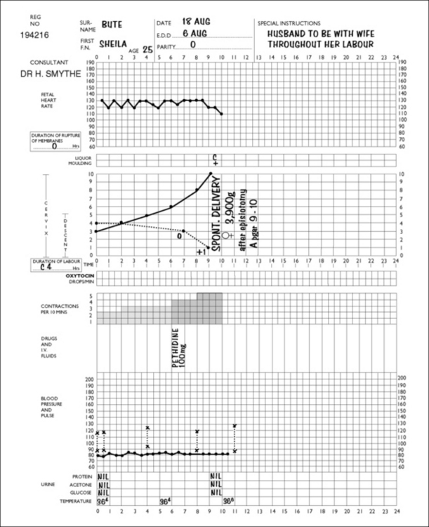
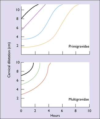
On admission to the delivery unit, the diagnosis of labour is either confirmed or rejected. If the woman is in labour, the time of admission is designated as the start of labour. A partogram is started and the progress of labour is marked on it (Fig. 8.1). Vaginal examinations are made every 2–4 hours and the cervical dilatation is recorded on the partogram. Action lines, printed on transparent plastic, are superimposed on the partogram (Fig. 8.2). In the active stage of labour the slowest acceptable rate of cervical dilatation is 1 cm/h. If the cervical dilatation lies to the right of the appropriate action line the membranes are ruptured and an incremental dilute oxytocin infusion is established in nulliparous women (and some multiparous women), provided that a single fetus presents as a vertex and there are no signs of fetal distress.

Fig. 8.2 Action lines for the partogram (Studd stencil). The lines show the slowest acceptable rate of cervical dilatation, commencing with the degree of dilatation on admission to the delivery unit.
Progress in the second stage of labour is judged by the descent of the fetal head and its rotation. One hour is allowed for the fetal head to reach the pelvic floor (the first or passive phase) and a further hour for the birth to be completed (the second or active phase). If there is delay in the passive phase then oxytocic augmentation is instituted. Delay in the active phase dictates close fetal monitoring and, where indicated, an operative vaginal delivery. Proponents of actively managed labour claim that the incidence of a labour lasting more than 12 hours is less than 3%; the caesarean section rate is 7–12% and the forceps rate is 8%. Opponents point out that only three randomized studies have been carried out, and that in these studies there was no reduction in the caesarean section or forceps rate.
Elective caesarean section
A small but increasing number of women, particularly those over the age of 35, inform their obstetrician during pregnancy that they wish to be delivered by caesarean section. The obstetrician should listen to the woman and discern the underlying reasons for her request. For some it may be a fear of the pain of labour, for others to avoid any risk of pelvic floor damage during delivery, or a perception that abdominal delivery removes all risks for the baby. It is particularly important that the risks of caesarean versus vaginal delivery are carefully explained (Box 8.3). If the woman confirms that she wishes to be delivered by caesarean section, the obstetrician should either agree to her wish or arrange for a consultation with another colleague.
Box 8.3 Effects of caesarean section compared with vaginal birth
Adapted from Caesarean Section, National Collaborating Centre for Women’s and Children’s Health. 2004. RCOG Press www.rcog.org.uk. Full details of absolute and relative risks can be obtained from full guideline.
Increased | No difference | Decreased |
---|---|---|
Abdominal pain | Blood loss >1000 ml | Perineal pain |
Bladder and ureteric injury | Infection-wound or endometritis | Urinary incontinence |
Need for laparotomy or D&C | Genital tract injury | Uterovaginal prolapse |
Hysterectomy | Back pain | |
ICU admission | Dyspareunia | |
Thromboembolism | Postnatal depression | |
Length of hospital stay | Neonatal morbidity (excluding breech) | |
Readmission | Neonatal intracranial haemorrhage | |
Placenta praevia | Brachial plexus palsy | |
Uterine rupture | Cerebral palsy | |
Maternal death | ||
Stillbirth in future pregnancies | ||
Secondary infertility | ||
Neonatal respiratory morbidity |
Home birth
Women choosing this type of delivery must receive good prenatal care; must be screened for any medical or obstetric abnormality; must accept transferral to a hospital if an abnormality arises during pregnancy or childbirth; and must have an experienced midwife. Even then home birth is less safe than giving birth in a midwife-controlled birthing centre.
ONSET OF LABOUR
In the weeks before labour starts the painless uterine contractions, which have been becoming increasingly frequent, merge into a prodromal stage of labour which may last up to 4 weeks. During this time the lower uterine segment expands to take the fetal head, which enters the upper pelvis. This relieves the pressure on the upper part of the abdomen (‘lightening’) but increases the pressure in the pelvis. Consequently, constipation and urinary frequency become apparent, and some patients complain of increased pressure in the pelvis and an increased mucoid vaginal discharge.
False labour
As term approaches many women complain of painful uterine contractions, which may seem to indicate the onset of labour. However, despite the contractions, progressive dilatation of the cervix fails to occur – this condition is termed false labour.
In it, the triple descending gradient of uterine activity fails to become established. A reverse gradient of uterine activity is present, the lower part of the uterus contracting nearly as strongly as the upper part. Because of this, cervical dilatation fails to occur and the pain of the uterine contractions is often felt as low backache.
Clinically, the painful contractions occur more often at night, but their frequency and intensity do not increase as time passes. A woman who complains of this pattern of uterine activity needs an explanation and, if the pains are distressing, requires treatment with analgesics, and perhaps a hypnotic so that she can enjoy a good night’s sleep.
Often the pains of false labour recur on a number of days and, in some cases, the reverse gradient of activity changes and true labour starts.
Onset of true labour
True labour may start and progress rapidly, or the start may be slow, with contractions only occurring at long intervals – the so-called sluggish uterus, or, in a more modern idiom, prolongation of the quiet (latent) phase of labour.
If the woman is becoming distressed that labour is not progressing, a more effective pattern of uterine activity can be obtained by performing an amniotomy and by starting an intravenous infusion of oxytocin. This regimen should only be instituted if there is no cephalopelvic disproportion and if the cervix is partly or wholly effaced, 2 cm dilated and soft.
The onset of labour is difficult to time with any degree of accuracy, and may be heralded by the following signs:
The transition into labour is gradual, but labour may be said to have begun when the cervix is at least 2 cm dilated and contractions become painful and regular, with diminishing intervals between each one.
Because of the difficulty in establishing the time of onset of labour with any degree of accuracy, many obstetricians mark its onset from the time the woman is admitted to hospital. This has advantages if the graphic method of recording the progress of labour (the partogram) is adopted.
Duration of labour
The duration of labour is not easy to determine precisely as its onset is often indefinite and subjective (Box 8.4). In studies of informed women whose labour started spontaneously there was a wide variation in the duration of labour, as can be seen in Table 8.1.
Several factors influence the duration of labour. These include the age of the woman, her parity, her knowledge of the process of childbirth, and the size of the fetus and its position in the uterus. Labour seems to last longer in nulliparous women, particularly older primigravidae, and if the baby presents in the occipitoposterior position.
Table 8.1 Duration (and range) of labour (in hours)
NULLIPARAE | MULTIPARAE | |
---|---|---|
1st stage | 8.25 (2–12) | 5.5 (1–9) |
2nd stage | 1 (0.25–1.5) | 0.25 (0–0.75) |
3rd stage | 0.25 (0–1)9.5 (2.25–14) | 0.25 (0–0.5)6 (1–10.25) |
Labour is usually shorter when the patient knows something of the physiology of normal labour, is in good physical and mental health at the onset of labour, and has confidence in her attendants. Delivery within 16 hours may be expected in 90% of nulliparous women (para 0), and the same proportion of multiparous women (para 1+) will deliver within 12 hours.
PROGRESS AND MANAGEMENT OF LABOUR AND BIRTH
The management of labour begins when the woman seeks admission to hospital, which she does when she believes or knows that she is in labour. As labour is a time of anxiety and stress, the attitude of the member of staff who admits her is most important. The care of the woman during her labour and childbirth may be conducted by a midwife (see also p. 66), or by midwives in partnership with an obstetrician or a general practitioner.
Admission
On admission the woman’s antenatal records are obtained and scrutinized for any past medical or obstetric problems, to check the history of the current pregnancy, and to make sure that the appropriate laboratory tests have been made. A history of the present labour is obtained, the frequency and strength of the uterine contractions are noted, and information is obtained about a ‘show’ of blood or mucus and whether or not the membranes have broken.
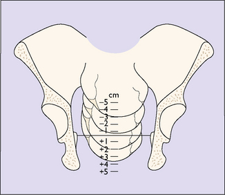
A general examination is made by a midwife or a doctor, the blood pressure, pulse and temperature being recorded. The abdomen is palpated to determine the presentation of the fetus and the position of the presenting part in relation to the pelvic brim (see Fig. 6.17). A vaginal examination may be carried out, with aseptic precautions, to determine the effacement and dilatation of the cervix and the position and station of the presenting part. The station of the presenting part is the level of the lowest fetal bony part (head or breech) in relation to an imaginary line joining the mother’s ischial spines. It is measured in centimetres above or below the ischial spines (Fig. 8.3). If the amniotic sac (the membranes) has ruptured this is noted. Evidence does not support the common practice of performing a routine 20-minute cardiotocogram (CTG) on admission in low-risk women. The ‘admission CTG’ is associated with higher intervention rates, i.e. augmentation of labour, epidural analgesia and operative delivery, without a clear improvement in neonatal outcome. The woman is transferred from the admission room to a bed in a delivery room (if she has not already been admitted to it) and a partogram is started which shows the progress of labour at a glance.

Full access? Get Clinical Tree

