Chapter 570 Congenital Adrenal Hyperplasia and Related Disorders
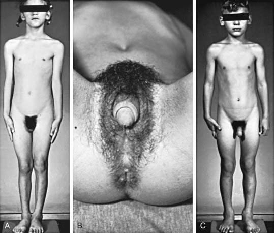
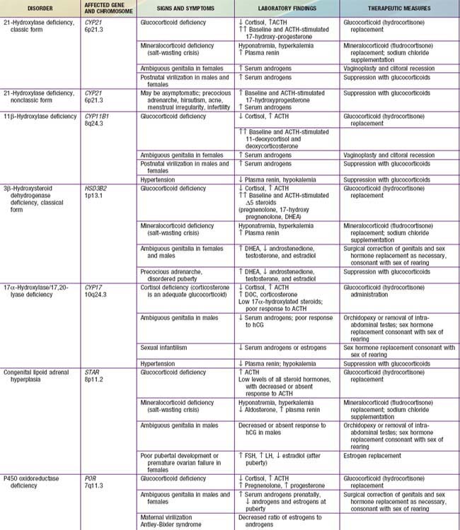
Congenital adrenal hyperplasia (CAH) is a family of autosomal recessive disorders of cortisol biosynthesis (normal adrenal steroidogenesis is discussed in Chapter 568). Cortisol deficiency increases secretion of corticotropin (ACTH), which in turn leads to adrenocortical hyperplasia and overproduction of intermediate metabolites. Depending on the enzymatic step that is deficient, there may be signs, symptoms, and laboratory findings of mineralocorticoid deficiency or excess; incomplete virilization or premature puberty in affected males; and virilization or sexual infantilism in affected females (Figs. 570-1 and 570-2 and Table 570-1).

Figure 570-1 A, A 6-yr-old girl with congenital virilizing adrenal hyperplasia. The height age was 8.5 yr, and the bone age was 13 yr. B, Notice the clitoral enlargement and labial fusion. C, Her 5-yr-old brother was not considered to be abnormal by the parents. The height age was 8 yr, and the bone age was 12.5 yr.

Figure 570-2 Three virilized females with untreated congenital adrenal hyperplasia. All were erroneously assigned male sex at birth, and each had a normal female sex-chromosome complement. Infants A and B had the salt-wasting form and received the diagnosis early in infancy. Infant C was referred at 1 yr of age because of bilateral cryptorchidism. Notice the completely penile urethra; such complete masculinization in females with adrenal hyperplasia is rare; most of these infants have the salt-wasting form.
570.1 Congenital Adrenal Hyperplasia Due to 21-Hydroxylase Deficiency
Etiology
More than 90% of congenital adrenal hyperplasia (CAH) cases are caused by 21-hydroxylase deficiency. This P450 enzyme (CYP21, P450c21) hydroxylates progesterone and 17-hydroxyprogesterone (17-OHP) to yield 11-deoxycorticosterone (DOC) and 11-deoxycortisol, respectively (see Fig. 568-1). These conversions are required for synthesis of aldosterone and cortisol, respectively. Both hormones are deficient in the most severe, “salt-wasting” form of the disease. Slightly less severely affected patients are able to synthesize adequate amounts of aldosterone but have elevated levels of androgens of adrenal origin; this is termed simple virilizing disease. These 2 forms are collectively termed classical 21-hydroxylase deficiency. Patients with nonclassical disease have relatively mildly elevated levels of androgens and may have signs of androgen excess after birth.
Epidemiology
Classical 21-hydroxylase deficiency occurs in about 1 in 15,000-20,000 births in most populations. Approximately 70% of affected infants have the salt-losing form, whereas 30% have the simple virilizing form of the disorder. In the USA, CAH is less common in African-Americans compared with white children (1 : 42,000 vs 1 : 15,500). Nonclassical disease has a prevalence of about 1 in 1,000 in the general population but occurs more frequently in specific ethnic groups such as Ashkenazi Jews and Hispanics.
Genetics
There are 2 steroid 21-hydroxylase genes—CYP21P (CYP21A1P, CYP21A) and CYP21 (CYP21A2, CYP21B)—which alternate in tandem with 2 genes for the 4th component of complement (C4A and C4B) in the human leukocyte antigen (HLA) major histocompatibility complex on chromosome 6p21.3 between the HLA-B and HLA-DR loci. Many other genes are located in this cluster. CYP21 is the active gene; CYP21P is 98% identical in DNA sequence to CYP21 but is a pseudogene due to 9 different mutations. More than 90% of mutations causing 21-hydroxylase deficiency are recombinations between CYP21 and CYP21P. Approximately 20% are deletions generated by unequal meiotic crossing-over between CYP21 and CYP21P, whereas the remainder is nonreciprocal transfers of deleterious mutations from CYP21P to CYP21, a phenomenon termed gene conversion.
The deleterious mutations in CYP21P have different effects on enzymatic activity when transferred to CYP21. Several mutations completely prevent synthesis of a functional protein, whereas others are missense mutations (they result in amino acid substitutions) that yield enzymes with 1-50% of normal activity. Disease severity correlates well with the mutations carried by an affected individual; for example, patients with salt-wasting disease usually carry mutations on both alleles that completely destroy enzymatic activity. Patients are frequently compound heterozygotes for different types of mutations (i.e., 1 allele is less severely affected than the other), in which case the severity of disease expression is largely determined by the activity of the less severely affected of the 2 alleles.
Pathogenesis and Clinical Manifestations
Aldosterone and Cortisol Deficiency
Because both cortisol and aldosterone require 21-hydroxylation for their synthesis, both hormones are deficient in the most severe, salt-wasting form of the disease. This form constitutes about 70% of cases of classical 21-hydroxylase deficiency. The signs and symptoms of cortisol and aldosterone deficiency, and the pathophysiology underlying them, are essentially those described in Chapter 569. These include progressive weight loss, anorexia, vomiting, dehydration, weakness, hypotension, hypoglycemia, hyponatremia, and hyperkalemia. These problems typically 1st develop in affected infants at approximately 10-14 days of age. Without treatment, shock, cardiac arrhythmias, and death may occur within days or weeks.
Prenatal Androgen Excess
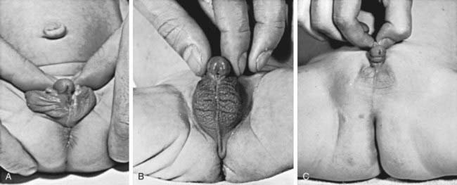
The most important problem caused by accumulation of steroid precursors is that 17-hydroxyprogesterone is shunted into the pathway for androgen biosynthesis, leading to high levels of androstenedione that are converted outside the adrenal gland to testosterone. This problem begins in affected fetuses by 8-10 wk of gestation and leads to abnormal genital development in females (see Figs. 570-1 and 570-2).
The external genitals of males and females normally appear identical early in gestation (Chapter 576). Affected females, who are exposed in utero to high levels of androgens of adrenal origin, have masculinized external genitalia (see Figs. 570-1 and 570-2). This is manifested by enlargement of the clitoris and by partial or complete labial fusion. The vagina usually has a common opening with the urethra (urogenital sinus). The clitoris may be so enlarged that it resembles a penis; because the urethra opens below this organ, some affected females may be mistakenly presumed to be males with hypospadias and cryptorchidism. The severity of virilization is usually greatest in females with the salt-losing form of 21-hydroxylase deficiency. The internal genital organs are normal, because affected females have normal ovaries and not testes and thus do not secrete antimüllerian hormone.
Male infants appear normal at birth. Thus, the diagnosis may not be made in boys until signs of adrenal insufficiency develop. Because patients with this condition can deteriorate quickly, infant boys are more likely to die than infant girls. For this reason, many states and countries have instituted newborn screening for this condition (see Newborn Screening, later).
Postnatal Androgen Excess
Untreated or inadequately treated children of both sexes develop additional signs of androgen excess after birth. Boys with the simple virilizing form of 21-hydroxylase deficiency often have delayed diagnosis because they appear normal and rarely develop adrenal insufficiency.
Signs of androgen excess include rapid somatic growth and accelerated skeletal maturation. Thus, affected patients are tall in childhood but premature closure of the epiphyses causes growth to stop relatively early, and adult stature is stunted (see Fig. 570-1). Muscular development may be excessive. Pubic and axillary hair may appear; and acne and a deep voice may develop. The penis, scrotum, and prostate may become enlarged in affected boys; however, the testes are usually prepubertal in size so that they appear relatively small in contrast to the enlarged penis. Occasionally, ectopic adrenocortical cells in the testes of patients become hyperplastic similarly to the adrenal glands, producing testicular adrenal rest tumors (Chapter 578). The clitoris may become further enlarged in affected females (see Fig. 570-1). Although the internal genital structures are female, breast development and menstruation may not occur unless the excessive production of androgens is suppressed by adequate treatment.
Adrenomedullary Dysfunction
Development of the adrenal medulla requires exposure to the extremely high cortisol levels normally present within the adrenal gland. Thus patients with classic CAH have abnormal adrenomedullary function, as evidenced by blunted epinephrine responses, decreased blood glucose and lower heart rates with exercise. Ability to exercise is unimpaired and the clinical significance of these findings is uncertain. Adrenomedullary dysfunction may exacerbate the cardiovascular effects of cortisol deficiency in untreated or undertreated patients.
Laboratory Findings (See Table 570-1)
Diagnosis of 21-hydroxylase deficiency is most reliably established by measuring 17-hydroxyprogesterone before and 30 or 60 min after an intravenous bolus of 0.125-0.25 mg of cosyntropin (ACTH 1-24). Nomograms exist that readily distinguish normals and patients with nonclassical and classical 21-hydroxylase deficiency. Heterozygous carriers of this autosomal recessive disorder tend to have higher ACTH-stimulated 17-hydroxyprogesterone levels than genetically unaffected individuals, but there is significant overlap between subjects in these 2 categories. However, in infants with frank electrolyte abnormalities or circulatory instability, it may not be possible or necessary to delay treatment to perform this test, as levels of precursors will be sufficiently elevated on a random blood sample to make the diagnosis.
Differential Diagnosis
Intersex conditions are discussed more generally in Chapter 582. The initial step in evaluating an infant with ambiguous genitals is a thorough physical examination to define the anatomy of the genitals, locate the urethral meatus, palpate the scrotum or labia and the inguinal regions for testes (palpable gonads almost always indicate the presence of testicular tissue and thus that the infant is a genetic male), and look for any other anatomic abnormalities. Ultrasonography is helpful in demonstrating the presence or absence of a uterus and can often locate the gonads. A rapid karyotype (such as fluorescence in situ hybridization of interphase nuclei for X and Y chromosomes) can quickly determine the genetic sex of the infant. These results are all likely to be available before the results of hormonal testing and together allow the clinical team to advise the parents as to the genetic sex of the infant and the anatomy of internal reproductive structures. Injection of contrast medium into the urogenital sinus of female pseudohermaphrodites demonstrates a vagina and uterus, and most surgeons utilize this information to formulate a plan for surgical management.
Prenatal Diagnosis
Prenatal diagnosis of 21-hydroxylase is possible late in the 1st trimester by analysis of DNA obtained by chorionic villus sampling or during the 2nd trimester by amniocentesis. This is usually done because the parents already have an affected child. Most often, the CYP21 gene is analyzed for frequently occurring mutations; more rare mutations may be detected by DNA sequencing.
Newborn Screening
Because 21-hydroxylase deficiency is often undiagnosed in affected males until they have severe adrenal insufficiency, all states in the USA and many other countries have instituted newborn screening programs. These programs analyze 17-hydroxyprogesterone levels in dried blood obtained by heel-stick and absorbed on filter paper cards; the same cards are screened in parallel for other congenital conditions such as hypothyroidism and phenylketonuria. Potentially affected infants are typically quickly recalled for additional testing (electrolytes and repeat 17-hydroxyprogesterone determination) at approximately 2 wk of age. Infants with salt-wasting disease often have abnormal electrolytes by this age but are usually not severely ill. Thus, screening programs are effective in preventing many cases of adrenal crisis in affected males. The nonclassical form of the disease is not reliably detected by newborn screening, but this is of little clinical significance because adrenal insufficiency does not occur in this type of 21-hydroxylase deficiency.
The main difficulty with current newborn screening programs is that to reliably detect all affected infants, the cutoff 17-hydroxyprogesterone levels for recalls are set so low that there is a very high frequency of false-positive results (i.e., the test has a low positive predictive value of approximately 1%). This problem is worst in premature infants. Positive predictive value can be improved by using cutoff levels based on gestational age, and by utilizing more specific 2nd-tier screening methods such as liquid chromatography followed by tandem mass spectrometry (LC-MS/MS).
Treatment
Glucocorticoid Replacement
Cortisol deficiency is treated with glucocorticoids. Treatment also suppresses excessive production of androgens by the adrenal cortex and thus minimizes problems such as excessive growth and skeletal maturation and virilization. This often requires larger glucocorticoid doses than are needed in other forms of adrenal insufficiency, typically 15-20 mg/m2/24 hr of hydrocortisone daily administered orally in 3 divided doses. Affected infants usually require dosing at the high end of this range. Double or triple doses are indicated during periods of stress,

Stay updated, free articles. Join our Telegram channel

Full access? Get Clinical Tree

