CHAPTER 3 Clinical examination of the patient with brachial plexus palsy
Summary box
Introduction
In this chapter, the physical examination of brachial plexus injury will be described, along with comments on how to integrate abnormal findings into one’s thought process regarding lesion localization and diagnosis. The technique for examining muscles innervated by direct branches of the brachial plexus will be discussed in the text and photo-illustrated. Examination techniques for muscles innervated by the major terminal branches of the brachial plexus (ie, median, ulnar, musculocutaneous, and radial nerves) will not be reviewed in the text, but will be described in the video presentation associated with this chapter. Other sources may be reviewed for additional detail regarding upper extremity motor testing.1,2,3
Visual inspection and general assessment
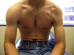
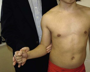
The patient should disrobe above the waist. Women are instructed to wear a sports bra so that they do not have to wear a gown during the examination, as a gown limits global comparison of the affected versus non-affected sides (Figure 3.1). Shoulder position in relation to the normal shoulder may indicate a trapezius palsy. Scapular winging at rest should be documented. Any atrophy is noted, both from an anterior and posterior view. Previous surgical scars and penetrating wounds, healed and unhealed, are examined. Trophic skin changes can result from nerve injury; they can present as ulcers or wounds on the hand and often heal very poorly. The paraspinal muscles, supraclavicular space, infraclavicular space, axilla, and upper arm are all inspected and palpated. Palpation begins in a gentle manner and progresses to a more deep assessment of the soft tissues. The course of the brachial plexus is tapped by a reflex hammer or the examiner’s fingers to check for Hoffman-Tinel’s sign. The pupils should be examined to exclude Horner’s syndrome, which would indicate a very proximal injury to the T1 spinal nerve, usually its avulsion from the spinal cord. Pulses in the extremities are assessed, and auscultation and percussion of the lungs to exclude paralysis of a hemidiaphragm secondary to a phrenic nerve palsy.
Muscle examination techniques TENTTMuscle (see video)
Documentation of motor strength requires an easy to remember and readily applicable grading scale. Use of a standardized scale is paramount for documenting a patient’s examination over time, as well as for comparing different individuals before and after treatment. The Medical Research Council published the classic motor function grading scale (MRC grading scale) shortly after World War II.1 This grading scale is presented in Table 3.1, and ranges from 5 (full strength) to 0 (no muscle contraction). The MRC grading scale is the best known and most utilized throughout the world. The simplicity of the MRC grading scale no doubt has contributed to its popularity; however, it has certain limitations that should be recognized. For example, it does not take into consideration range of motion, tends to be weighted toward weaker contractions, and is poorly applicable to certain muscles (e.g., rhomboids). In response to these deficiencies, other grading scales have been described. The Louisiana State University (LSU) motor function grading scales are popular amongst peripheral nerve surgeons and are specific for each major peripheral nerve or element of the brachial plexus.4
Table 3.1 Medical Research Council motor function grading scale
| Grade | Function |
|---|---|
| 5 | Full strength |
| 4 | Movement against resistance |
| 3 | Movement against gravity only |
| 2 | Movement with gravity eliminated |
| 1 | Muscle contraction but no movement |
| 0 | No muscle contraction |
This section presents the examination technique for muscles innervated by direct branches of the brachial plexus. Examination techniques for other distal upper extremity musculature are well-known, are presented elsewhere in detail,1,3,4 and are illustrated in the video supplement to this chapter.
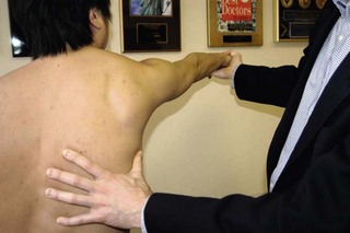
The long thoracic nerve is a proximal branch of the brachial plexus, arising from the proximal C5, C6, and C7 spinal nerves, that innervates the serratus anterior muscle. This muscle originates on the lateral surfaces of the upper 8 ribs and inserts on the entire medial border of the scapula. It pulls the scapula away from the midline and forward around the thorax (scapular abduction). It also rotates the lateral angle of the scapula upward. Most importantly, however, this muscle fixes and stabilizes the scapula so that muscles originating from it can function properly. Furthermore, anterior arm flexion is stabilized by the serratus anterior, with flexion above 90 degrees mostly due to upward rotation of the scapula. Injury to the long thoracic nerve causes winging of the scapula. Winging may occur at rest, is most noticeable at the inferomedial angle, and is classically worsened when the patient pushes forward against resistance. Winging continues when the upper extremity is locked in extension with the shoulder girdle protracted forward (anteriorly). To test this muscle, instruct patients to reach for a point on the wall in front of them, and then apply resistance at the hand or wrist while stabilizing the thorax with the other hand (Figure 3.2). A common mistake is to not have patients displace their shoulder girdle far enough forward, because without doing so, scapular winging due to trapezius or rhomboid weakness may be misdiagnosed as a long thoracic palsy. It is important to note that all 3 causes of scapular winging cause winging when the arm is pushed against resistance across the chest with the arm bent. Only serratus anterior weakness will show severe winging when pushing the fully extended arm forward (protracting) against resistance (Figure 3.3).
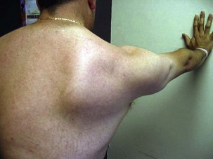
Figure 3.3 Scapular winging is present on the patient’s right side secondary to a spinal accessory palsy.
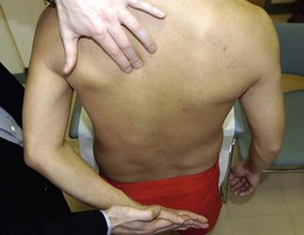
The dorsal scapular nerve is a very proximal branch from the C5 spinal root. This nerve innervates both the major and minor rhomboid muscles. The rhomboids connect the medial edge of the scapulae to the spinal column. When contracted, the rhomboids pull the scapula toward the midline (scapular adduction and retraction) and superiorly (downward rotation of the lateral angle). The rhomboids move the scapula in the opposite direction to that of the serratus anterior muscles. With chronic denervation, wasting of this muscle deep to the trapezius is evident. With rhomboid weakness, there may be mild scapular winging at rest, especially at the inferior medial edge. The scapula may also be displaced laterally and inferiorly and rotated laterally. To test the rhomboids, have patients place their palm facing outward on their lower back. Instruct them to push the palm away from the lower back as the examiner applies resistance to the hand as well as to the arm (the arm is pushed anterolaterally around the thorax) (Figure 3.4). The rhomboids are observed and palpated along the medial scapular border during this maneuver. If the rhomboids are weak, only resist at the hand. An alternate method to examine the rhomboids is to have patients bring their shoulders and scapulae together posteriorly. In this position, the contracted rhomboids can be palpated between the lower aspects of the scapulae. The dorsal scapular nerve can also provide partial innervation to the levator scapulae as it passes underneath this muscle. This muscle assists the upper trapezius in shrugging the shoulders.
The suprascapular nerve (C5, C6) originates from the upper trunk of the brachial plexus and passes the inferior belly of the omohyoid to the suprascapular notch through which it passes to the posterior surface of the scapula. The suprascapular nerve innervates the supraspinatus and infraspinatus muscles. The supraspinatus attaches to the superior aspect of the humeral head and mediates the initial 20-30 degrees of arm abduction. The infraspinatus attaches to the posterior lateral aspect of the humeral head and is the primary external rotator of the arm. Test the supraspinatus muscle by having patients abduct a straight arm from their side against resistance (Figure 3.5). Test the infraspinatus muscle by having patients flex their forearm to 90 degrees, and while stabilizing their elbow against their side, instruct them to externally rotate their arm against resistance, like a tennis swing (Figure 3.6). Contraction can be observed and palpated over the scapula while testing these muscles. With chronic denervation, atrophy above (supraspinatus) or below (infraspinatus) the scapular spine is readily appreciated.
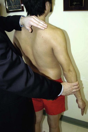
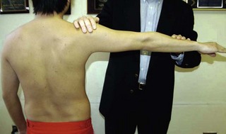
The axillary nerve (C5, C6) arises from the posterior cord deep to the axillary artery and divides into an anterior and posterior division near the humeral neck as it passes medially and posteriorly to it. The anterior division innervates the anterior and lateral deltoid. The posterior division gives a branch to the teres minor, innervates the posterior portion of the deltoid, and gives a sensory branch to the lateral shoulder region. The teres minor assists the infraspinatus in externally rotating the arm. It also weakly assists the teres major in adducting an extended arm. It is not possible to test this muscle in complete isolation, but one may observe and palpate it if the patient is thin. The deltoid is the prime abductor, as well as flexor (lifting the arm in front of the body) of the arm. The initial 30 degrees of abduction is primarily controlled by the supraspinatus, whereas abduction above 90 degrees has an important trapezius and serratus component that tilts the shoulder girdle upward. Therefore, test the deltoid by having patients abduct their arm between 30 and 90 degrees against resistance (Figure 3.7). The deltoid has 3 separate heads: the anterior, lateral, and posterior. Abducting the arm to the side and slightly in front of the body tests the anterior and lateral heads of the deltoid. To assess the posterior head, have patients place a straightened arm at almost 90 degrees abducted, and then ask them to move the arm posteriorly and superiorly against resistance (Figure 3.8).
< div class='tao-gold-member'>
Stay updated, free articles. Join our Telegram channel

Full access? Get Clinical Tree


