Chronic Severe Respiratory Insufficiency

Chapter 412 Chronic Severe Respiratory Insufficiency


Improvements in the treatment of acute respiratory failure and advancements in invasive and noninvasive ventilation have led to an increase in the number of pediatric patients receiving long-term mechanical ventilation. Infants, children, and adolescents with disorders of central control of breathing, disease of the airways, residual lung disease after severe respiratory illness, and neuromuscular disorders may experience hypercarbic and/or hypoxemic chronic respiratory failure. Although it is generally possible to identify a primary cause for the respiratory failure, many children have multiple causative factors. Chronic respiratory failure is pulmonary insufficiency for a protracted period, usually 28 days or longer. Patients are maintained on long-term ventilation until they recover from the initial pulmonary insult. Patients with conditions such as central hypoventilation, progressive neuromuscular disease, and high quadriplegia may need ventilatory support indefinitely. The preferred site for a patient’s care after initial discharge with a ventilator is within the family home. When social circumstances do not allow this placement, patients may be placed in a highly skilled nursing facility.


Less than 1% of patients admitted to pediatric intensive care units require long-term noninvasive or invasive ventilatory assistance. A survey from Massachusetts by Graham, Fleegler, and Robinson (2007) identified 197 children undergoing long-term ventilatory support, a threefold increase over a 15-year period. The majority of the primary diagnoses (54%) were congenital or perinatal-acquired neurologic or neuromuscular disorders. Chronic lung disease of prematurity represented only 7% of the sample, a significant shift downward, presumably a result of improvement in neonatal care over the same period. Seventy percent of the patients were cared for at home.


412.1 Neuromuscular Diseases


Neuromuscular diseases (NMDs) of childhood include muscular dystrophies, metabolic and congenital myopathies, anterior horn cell disorders, peripheral neuropathies, and diseases that affect the neuromuscular junction. Decreases in muscle strength and endurance resulting from neuromuscular disorders can affect any skeletal muscle, including muscles involved in respiratory function. Of particular concern are those muscles mediating upper airway patency, generation of cough, and lung inflation. Acute respiratory insufficiency is often the most prominent clinical manifestation of several acute neuromuscular disorders, such as high-level spinal cord injury, poliomyelitis, Guillain-Barré syndrome (Chapter 608), and botulism (Chapter 202). Although much more insidious in its clinical course, respiratory dysfunction constitutes the leading cause of morbidity and mortality in progressive neuromuscular disorders (e.g., Duchenne muscular dystrophy [Chapter 601], spinal muscular atrophy, congenital myotonic dystrophy, myasthenia gravis [Chapter 604], and Charcot-Marie-Tooth disease [Chapter 605]).


Two of the most common and best understood NMDs of childhood are Duchenne muscular dystrophy (DMD) and spinal muscular atrophy (SMA). Principles utilized to evaluate and treat respiratory insufficiency in these disorders are often based on medical consensus rather than research. Despite this limitation, these principles are useful in the management of DMD and SMA as well as other less common NMDs.


Treatment


Even though gene-targeted therapies are being developed for some NMDs, current interventions are primarily supportive rather than curative. Close surveillance through periodic review of the history and physical examination is critical. The development of personality changes, such as irritability, decreased attention span, fatigue, and somnolence, may point to the presence of sleep-associated gas exchange abnormalities and sleep fragmentation. Changes in speech and voice characteristics, nasal flaring, and the use of other accessory muscles during quiet breathing at rest may provide sensitive indicators of progressive muscle dysfunction and respiratory compromise. Although the frequency of periodic re-evaluation needs to be tailored to the individual patient, tentative guidelines were developed for patients with DMD; an abbreviated summary of such recommendations, applicable to all children with NMDs, is provided in Table 412-1.


Table 412-1 PROPOSED GUIDELINES FOR INITIAL EVALUATION AND FOLLOW-UP OF PATIENTS WITH NEUROMUSCULAR DISEASE


INITIAL EVALUATION BASIC INTERVENTION/TRAINING
History/physical/anthropometrics Nutritional consultation and guidance
Lung function and maximal respiratory pressures (PFTs) Regular chest physiotherapy
Arterial blood gases Use of percussive devices
Polysomnography* Respiratory muscle training
Exercise testing (in selected cases) Annual influenza vaccine
If vital capacity >60% predicted or maximal respiratory pressures >60 cm H2O Evaluate PFTs every 6 mo
CXR and polysomnography every year
If vital capacity <60% predicted or maximal respiratory pressures <60 cm H2O Evaluate PFTs every 3-4 mo
CXR, MIP/MEP every 6 mo
Polysomnography every 6 mo to every year

CXR, chest x-ray; MEP, maximal expiratory pressure; MIP, maximal inspiratory pressure; PFT, pulmonary function test.


* Please note that if polysomnography is not readily available, multichannel recordings including oronasal airflow, nocturnal oximetry, and end-tidal carbon dioxide levels may provide an adequate alternative.


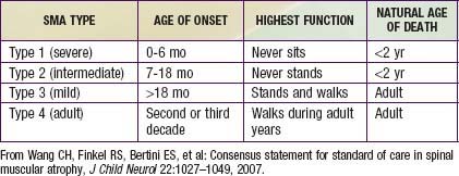
Guidelines for evaluation and management of patients with SMA were developed on the basis of expert consensus. Four classifications of SMA, based on age of onset and level of function, are recognized (Table 412-2). Treatment of SMA is focused on level of function (non-sitter, sitter, or walker) rather than SMA type. Unlike patients with DMD, patients with SMA do not demonstrate correlation between pulmonary function and need for mechanical ventilatory support. Rather, longitudinal monitoring for signs and symptoms of sleep-disordered breathing and ineffective airway clearance should be utilized to direct patient care.


412.2 Congenital Central Hypoventilation Syndrome


Congenital central hypoventilation syndrome (CCHS) is a clinically complex disorder of respiratory and autonomic regulation. In the classic case of CCHS, symptoms of alveolar hypoventilation are manifest during sleep only, but in more severe cases, the symptoms are manifest during sleep and wakefulness. The classic syndrome is further characterized by ventilatory failure to respond to hypercarbia and hypoxemia during wakefulness and sleep as well as physiologic and/or anatomic autonomic nervous system (ANS) dysregulation (ANSD). The physiologic ANSD may include all organ systems affected by the ANS, specifically the respiratory, cardiac, sudomotor, ophthalmologic, neurologic, enteric systems, and more. The anatomic or structural ANSD in CCHS includes Hirschsprung disease and tumors of neural crest origin (neuroblastoma, ganglioneuroma, or ganglioneuroblastoma). Most patients with CCHS present in the neonatal period, although later-onset CCHS (LO-CCHS) may manifest in infancy, childhood, and even adulthood. The initial symptoms include diminished tidal volume and a typically monotonous respiratory rate with cyanosis and hypercarbia. Diagnosis and management of children with CCHS have improved considerably, owing to greater knowledge in genetic testing, comprehensive care, and availability of technology for the home.


Genetics


In 2003, the paired-like homeobox 2B (PHOX2B) gene was identified as the disease-defining gene for CCHS. This gene, which is essential to the embryologic development of the ANS from the neural crest, is expressed in key regions that explain much of the CCHS phenotype. Individuals with CCHS are heterozygous for either a polyalanine repeat expansion mutation (PARM) in exon 3 of the PHOX2B gene (normal number of alanines is 20 with normal genotype 20/20), such that individuals with CCHS have 24-33 alanines on the affected allele (genotype range is 20/24-20/33), or a non–polyalanine repeat expansion mutation (NPARM) resulting from a missense, nonsense, or frameshift mutation. Roughly 90% of the cases of CCHS have PARMs and the remaining ≈10% of cases have NPARMs. The specific type of PHOX2B mutation is clinically significant as it can help with anticipatory guidance in patient management.


The majority of CCHS cases occur because of a de novo PHOX2B mutation, but 5-10% of children with CCHS inherit the mutation from an asymptomatic parent who is mosaic for the PHOX2B mutation. CCHS is inherited in an autosomal dominant manner. Therefore, an individual with CCHS has a 50% chance of transmitting the mutation, and resulting disease phenotype, to each child. Mosaic parents have up to a 50% chance of transmitting the PHOX2B mutation to each offspring. Genetic counseling is essential for family planning and for preparedness in the delivery room for an anticipated CCHS birth. PHOX2B testing is advised for both parents of a child with CCHS to anticipate risk of recurrence in subsequent pregnancies. Further, prenatal testing for PHOX2B mutation is clinically available (www.genetests.org) for families with a known PHOX2B mutation.


Neuropathology


Anatomic findings in the brains of individuals with CCHS from early MRI studies were unremarkable, and those from autopsies were inconsistent. In a small cohort of adolescents with suspected CCHS, though without PHOX2B mutation confirmation, neuropathologic brainstem changes were identified by diffusion tensor imaging (DTI) in structures known to mediate central chemosensitivity and to link a network of cardiovascular, respiratory, and affective responses. The neuroanatomic defects in CCHS are likely the result of focal PHOX2B (mis)expression coupled with, in the suboptimally managed patient, sequelae of recurrent hypoxemia/hypercarbia. On the basis of rodent studies and functional MRI (fMRI) in humans, the following regions pertinent to respiratory control show PHOX2B expression in the pons and medulla of the brainstem: locus coeruleus, dorsal respiratory group, nucleus ambiguus, parafacial respiratory group, among other areas. Physiologic evidence suggests that the respiratory failure in these children is mostly based on defects in central mechanisms, but peripheral mechanisms (mainly carotid bodies) are also important.


Patients with CCHS have deficient carbon dioxide sensitivity during wakefulness and sleep; they do not respond with increased ventilation or arousal to hypercarbia during sleep. During wakefulness, a subset of patients may respond sufficiently to avoid hypercarbia, but most individuals with CCHS have hypoventilation that is severe enough that hypercarbia is apparent in the resting awake state. Children with CCHS also have altered sensitivity to hypoxia while awake and asleep. A key feature of CCHS is the lack of respiratory distress or sense of asphyxia with physiologic compromise. This lack of responsiveness to hypercarbia and/or hypoxemia with subsequent respiratory failure does not improve with advancing age. A subset of older children with CCHS may show an increase in ventilation (specifically increase in respiratory rate rather than increase in tidal volume) when they are exercised at various work rates, a response that is possibly secondary to neural reflexes from rhythmic limb movements—although the increase in minute ventilation is often insufficient to avoid physiologic compromise.


Clinical Manifestations


Patients with CCHS usually present in the first few hours after birth. Most children are the products of uneventful pregnancies and are term infants with appropriate weight for gestational age; Apgar scores have been variable. They do not show signs of respiratory distress, but their shallow respirations and respiratory pauses (apnea) evolve to respiratory failure with apparent cyanosis in the first day of life. In neonates with CCHS, the PaCO2 accumulates during sleep to very high levels, sometimes >90 mm Hg, and may decline to normal levels after the infants awaken. This problem becomes most apparent with failure of multiple attempts at extubation in an intubated neonate (who appears well with ventilatory support but in whom respiratory failure develops after removal of the support). However, the more severely affected infants hypoventilate awake and asleep; thus the previously described difference in PaCO2 between states is not apparent. Often, the respiratory rate is higher in rapid eye movement (REM) sleep than in non-REM sleep in individuals with CCHS.

Stay updated, free articles. Join our Telegram channel

Jun 18, 2016 | Posted by in PEDIATRICS | Comments Off on Chronic Severe Respiratory Insufficiency

Full access? Get Clinical Tree

Get Clinical Tree app for offline access