Chronic Pelvic Pain


Chronic Pelvic Pain


John F. Steege


Chronic pelvic pain (CPP) is generally defined as pain present in the pelvis, more often than not, for 6 months or longer. It may be cyclic or continuous in nature. Pain that is present at least 5 days/month occurs in 12% of women of reproductive age and is the reason for hysterectomy in approximately 12% of such procedures.1,2 Approximately half of all operative laparoscopies are performed to investigate problems with pain, many of those chronic in nature.3,4 The surgically minded clinician may conclude that once any pathology is found, the source of the pain has been found as well. The point of this chapter is to suggest a more thoughtful consideration of pain as a disease rather than as solely a symptom of pathology.

As a multifactorial disease, CPP often challenges the best of clinicians. When organic pathology is detected, its precise relationship to the production of symptoms is often uncertain. The longer the pain has gone on, the more likely treatment will be prolonged and perhaps incompletely successful. For the surgically oriented gynecologist, this problem can be especially frustrating. Surgical solutions are not always apparent, and pain may persist despite a satisfactory anatomic result. The challenge for the gynecologist is to learn when traditional gynecologic medical and surgical treatments will work and when they are less likely to succeed.

The temptation to resort to surgery is inherent in gynecologic training. The few placebo-controlled trials of surgery that have been carried out have demonstrated substantial nonspecific (placebo) effects of surgery, which can occur in 65 to 100% of cases and may last as long as a year.5,6 The surgeon seeing the patient 4 to 6 weeks after surgery may witness partial or complete relief of pain, only to see it return 3 to 12 months later and assume this must represent a return of disease. The most egregious example of this from my own practice is a 39-year-old woman who, prior to my evaluation, had undergone 37 laparoscopies to treat her pelvic pain. Each had been followed by 3 to 6 months of pain relief, although minimal intervention was done each time; she still had all reproductive organs in place.

This review focuses on information about pain pathways that may help to explain some of these phenomena. It reviews basic elements of history and physical examination pertinent to the various diagnoses and sources of CPP. Current treatments for each disorder are summarized. More importantly, we also deal with the cases in which treatment of apparent pathology is unsuccessful in relieving pain.


PATHWAYS OF PAIN PERCEPTION

Put simply, clinical pain falls into two categories: (a) nociceptive pain, caused by true peripheral tissue damage or physiologic processes, and (b) centralized pain, sometimes called “neuropathic,” which continues with very little or no input from damaged peripheral tissues, and is presumed to be related to damage to or dysfunction of the peripheral or central nervous system (CNS). Treatments aimed at peripheral tissues, whether medical or surgical, are more likely to be helpful when the pain is truly nociceptive. Conversely, centralized/neuropathic pain will not respond to these measures (beyond the placebo effect) but will only yield to pharmacologic and other nonsurgical measures that target CNS processes.

Before describing the typical clinical hallmarks of these two basic types of pain, a review of pain neuro-physiology is in order. Nociceptive signals emerging from pelvic visceral structures travel cephalad through one or more of several routes.7 (“Nociceptive signals” are peripheral nerve impulses that are destined to produce negatively perceived sensations; “pain” is a brain interpretation of sensations indicative of real or potential tissue damage.) Sympathetic afferent signals arising from the uterus, cervix, and immediately adjacent portions of the uterosacral, broad, and round ligaments travel via the inferior and superior hypogastric plexuses to the sympathetic chain. From there, they go to the T10 to T12 levels of the spinal cord. Additional sensation may be provided through the nervi erigentes, or pelvic nerves, which cross the pelvic floor and ultimately join the S2 to S4 segments of the spinal cord. Fibers from both the sympathetic and parasympathetic systems traverse Frankenhäuser plexus and the uterovesical ganglion.

The gate control theory, developed by Melzack and Wall in the 1950s and early 1960s, provides a model to help explain some of the variability in clinical pain patterns.8 It is a useful model of integration
of physical and psychological processes into a single scheme. The major contribution of this theory was to suggest that higher centers in the brain have the capacity to physiologically down- or upregulate signals that approach the spinal cord. This regulation may determine the degree to which peripheral nociceptive signals travel cephalad to reach conscious perception.

Building on this theory, further neurophysiologic research has focused on these spinal cord mechanisms.9 Glial cells, once thought to have only a nutritive role for surrounding neurons, are now recognized as playing a major role in this modulation process.10 Uncontrolled activation of microglial cells under neuropathic pain conditions induces the release of proinflammatory cytokines (interleukin [IL]-1β, IL-6, tumor necrosis factor) and complement components that facilitate pain transmission. Microglial activation can lead to altered activity of opioid systems, and neuropathic pain is characterized by resistance to morphine.10

It is now known that there are several categories of narcotic receptor sites both in the brain and the spinal cord. Indeed, some inhibit pain perception, whereas others actually facilitate it, giving rise to narcotic-induced hyperalgesia. In addition, the spinal cord appears to have receptors for local anesthetics and numerous other neurotransmitter-like substances, including cannabinoids.

Many researchers in pain have found the gate theory too limiting as well. Melzack11 suggested the term “neuromatrix” to describe a weblike set of communications, ascending and descending, that are involved in nociception and ultimately, in the perception of pain. The model allows inclusion of multiple new factors that have been the foci of recent research on spinal cord and central elements of the pain perception system. For example, recent evidence has begun to highlight the role of neuroimmune activation following tissue injury as an important mechanism in the development of chronic pain via cytokine-mediated central sensitization of neural pathways.12

Clinically, centralized pain is typically present continuously, is intense, and often is described as burning in nature. The patient may demonstrate sympathetic activation manifested by tachycardia, hypertension, hyperhidrosis, and insomnia. When examined, she may demonstrate hyperalgesia (excessive response to noxious stimuli) and/or allodynia (painful response to nonpainful stimuli).

Spinal cord “wind-up” occurs when repeated, low-level nociceptive stimuli from the periphery elicit progressively more dramatic responses from second-order neurons at the spinal cord level even though the intensity of the stimuli may remain constant or even be diminished. This phenomenon has been well demonstrated in acute pain models at the animal level.13 There is considerable speculation regarding the impact of this process on chronic pain symptoms. When examining a patient with pain, the tendency for repeated examination to elicit pain from initially nontender areas is a phenomenon consistent with “wind-up.”

It has also long been observed that visceral organs and portions of the somatic system often share nerve supply from the same level of the spinal cord. Pain from viscera may thus be referred to the appropriate somatic dermatome, a process labeled viscerosomatic convergence. With time, however, stimuli applied to somatic structures may sometimes reproduce a nociceptive response perceived as though it emanated from the original offending viscus. For example, pain from a vaginal fornix in a posthysterectomy patient can be referred to a relatively localized area of the abdominal wall. Typically, this spot is about 5 cm cephalad to the inguinal ligament and 5 cm from the midline toward the symptomatic side. With time, this abdominal wall area may itself become intrinsically tender. Palpation of this spot may reproduce the vaginal apex pain. Interestingly, local anesthetic block of the abdominal wall tender spot (trigger point) can decrease the sensitivity of the vaginal apex.

There are complex interactions among the reproductive organs, the urinary tract, and the colon. One phenomenon associated with these interactions is viscerovisceral hyperalgesia. In this condition, inflammation or congestion in an organ (e.g., the reproductive organs) enhances pain in viscera, skin, or muscle that share common spinal cord segments.14, 15, 16

Although acute and chronic pain are quite different physiologic processes, some lessons learned from acute pain management apply well to the management of chronic pain. For example, studies regarding the impact of preemptive analgesia on acute postoperative pain suggest that the ideal postoperative pain regimen is multifaceted. The ideal regimen simultaneously interrupts the nociceptive signals at the peripheral tissue level, blocks receptor sites within the spinal cord, and alters the central perception of pain.17 In many chronic pain conditions, this would also be the ideal regimen. Treatments need to be applied at the peripheral tissue level and include pharmacologic measures directed toward spinal cord and central mechanisms as well.

All chronic pain is profoundly influenced by psychological processing and responses.18 Patients with identical diseases will vary greatly in pain severity, functional impairment, and pain behaviors.19 Operant conditioning factors20 and cognitive behavioral factors21 may be important in the maintenance of chronic pain behaviors.

Chronic pain is also influenced by psychosocial and psychiatric disturbances such as cultural influences, social support, comorbid mood disorder, and drug abuse.22 The contribution of psychological, social, and psychiatric factors should not lead to the conclusion that a pain syndrome is primarily psychogenic. Pain related
exclusively or primarily to psychological factors occurs but is far less prevalent than pain associated with organic processes that are powerfully influenced by psychosocial mediators and psychiatric comorbidities.


EVALUATION OF CHRONIC PAIN SYNDROMES


Physical Examination

The data provided by the history and observations made during the history taking process will provide focus for the physical examination. Before and during the history, the clinician may have the opportunity to observe posture, stance, gait, and sitting behavior. For example, a person with levator spasm will often sit forward on the chair and rest more of her weight on one buttock or the other. Sitting perfectly straight often aggravates the pain from this disorder.

The abdominal examination in a woman with CPP may be more informative if done in the following manner. Ask the patient to point to the area of pain with one finger and outline and circle the area involved. Ask her then to press as hard as she feels it is necessary in order to elicit the pain she experiences. Done in this manner, the impact of her anxiety about being examined may be diminished and a more accurate assessment of tissue sensitivity obtained. The patient is then asked to flex the abdominal muscles by raising only her head off the table and then asked to repeat the same palpation. If the pain elicited by palpation with the abdomen flexed is either the same or greater than the pain of the first palpation, then the abdominal wall itself is likely to be a source of the pain or a “pain generator.” This is a positive Carnett sign. If pain from palpation of the tensed abdominal wall is less, then an internal visceral source(s) is more likely.

The abdominal wall should then be systematically palpated by the examiner’s index finger in an effort to identify local spots of tenderness or “trigger points.” A trigger point is a hyperirritable spot within a taut band of skeletal muscle fibers that is painful on compression and may give rise to referred pain, tenderness, tightness, and sometimes, local twitch response and autonomic phenomena.
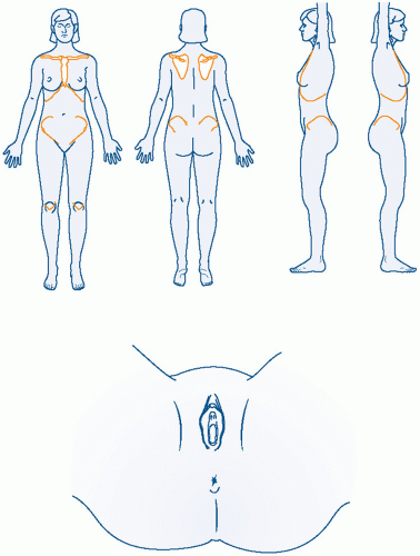
26,27 A diagrammatic record of the examination findings is valuable.

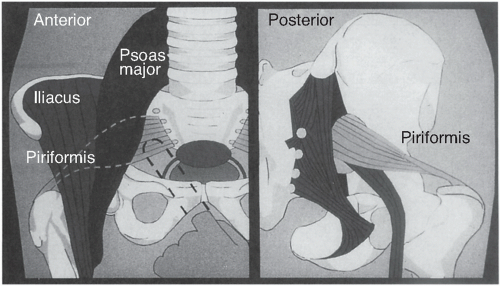
Specific elements of the musculoskeletal examination should be performed to look for piriformis spasm and/or psoas muscle pain. The piriformis originates at the lateral margin of the sacrum, traverses the greater sciatic
notch, and inserts on the greater trochanter of the femur. Its function is to externally rotate the thigh. It also forms a muscular bed for the sacral plexus of nerves in the pelvis, and some or all of the sciatic nerve goes through the belly of the piriformis in 25% of women. With her hip and her knee partially flexed (i.e., in the stirrups), ask the patient to externally rotate each thigh in turn against resistance. Pain in the posterior hip area with this maneuver suggests piriformis spasm. Irritation of the sciatic nerve by this spasm can cause hyperreflexia and “pseudosciatica” or pain that mimics sciatic nerve compression seen in lumbosacral disc disease.

In the lateral decubitus position, the hip is actively and passively extended and flexed. Pain with these maneuvers suggests a psoas muscle origin. Further elements of the musculoskeletal screening examination are discussed elsewhere (Table 7.2).23

The pelvic examination in the patient with CPP is more informative when performed in a careful stepwise fashion. Meticulous attention is paid to each potential contributing organ system. While collecting this information, the examiner should be thinking of making a list of contributing factors, not examining until “the cause” of the pain is found. First, place one index finger in the vaginal introitus and ask for voluntary contraction and relaxation. Inability to exert conscious control of the bulbocavernosus muscles suggests vaginismus. This diagnosis should, of course, be corroborated by additional history. The presence of good voluntary control during pelvic examination does not exclude the diagnosis of vaginismus that may occur during sexual situations.28

Extending the index finger beyond the bulbocavernosus muscle, one can usually palpate the levator muscles on the right and left sides at approximately the 4:30 and 7:30 positions. In the asymptomatic person, a sense of pressure is experienced. When levator spasm is present, gentle palpation on these muscles may reproduce the patient’s sense of pelvic pressure (a “falling out” sensation) and/or the pain of dyspareunia. Further verification is obtained when the symptom is exacerbated by voluntary contraction of the levators.


TABLE 7.2 Musculoskeletal Screening Examination for Chronic Pelvic Pain


Observation/Maneuver


Typical Abnormal Findings


Gait, stance


Scoliosis


Lateral standing view


One extremity externally rotated; exaggerated kyphosis-lordosis


Sitting


Raised up on one buttock (levator spasm)


With supine flexion of one hip, observe other hip (Thomas test)


Contralateral hip flexes, indicating hip flexor contracture


External rotation of the thigh against resistance (sitting, supine, or during pelvic examination)


Pain on external rotation indicates piriformis syndrome


Psoas flexion and extension


Limited extension and/or pain with flexion indicates psoas shortening ± spasm


Digital vaginal examination


Pain indicates levator spasm


The next step is to press directly on the coccyx, reaching it with the vaginal finger by going around the rectum on either side. External palpation of the coccyx combined with this maneuver will normally allow it to flex and extend through an approximately 30-degree angle. This is normally not a painful maneuver. If pain is present, it may be related to coccydynia or adjacent levator spasm.

The piriformis muscle can be tested by the maneuvers described earlier. When these maneuvers suggest piriformis spasm, this can be further confirmed on pelvic examination. The belly of the piriformis can be easily felt transvaginally when the thigh is externally rotated against resistance. When palpation of the piriformis muscle belly reproduces pain, then spasm is likely to be present. A false-positive interpretation of this maneuver can occur if intrinsic cul de sac disease is present because the vaginal examination finger must traverse this area to reach the piriformis muscle (Fig. 7.3).

The anterior vaginal wall, urethra, and bladder trigone should be examined either before or after the muscular examinations just described, depending on the clinical situation. In general, it is best to examine the (likely) nontender areas first. This technique helps minimize the tendency to experience pressure as pain (“wind-up”) once pain has been elicited. In many cases, the clinician can make the presumptive diagnosis of chronic urethritis, trigonitis, or interstitial cystitis on the basis of careful history combined with focused digital examination. If complex therapeutic interventions are being contemplated, then cystoscopic confirmation (i.e., cystoscopy with hydrodistention) should first be obtained. In the asymptomatic patient, pressure on the bladder base creates only urinary urgency without replicating the clinical pain.

Unimanual, single-digit transvaginal examination should continue by testing the intrinsic sensitivity of the cervix to palpation and traction. The adnexal areas are then examined in a similar manner, first unimanually and then adding the abdominal hand. Separating the pelvic exam into three discrete steps (unimanual vaginal exam alone, unimanual external exam of lower abdomen alone, and then examining the pelvis bimanually) helps distinguish internal visceral pain from pain originating from the abdominal wall.

Rather than following a rigid sequence, speculum examination should be done at a time when it makes the most sense, depending on the type of pain under consideration. For example, in the patient with a history suggesting posthysterectomy vaginal apex pain, it is probably wisest to complete the muscular examination as well as the adnexal palpation prior to insertion of the speculum. In this manner, the areas more likely to be
nontender are examined prior to the speculum directly touching the potentially sensitive vaginal apex.


FIGURE 7.3 Vaginal examination of piriformis muscle. (Modified from Barton PM. Piriformis syndrome: a rational approach to management. Pain. 1991; 47[3]:345-352.)

In contrast, if the history suggests an adnexal source of pain, then the pelvic examination should begin with the speculum examination (as is traditionally done). The speculum examination is then followed by a pelvic musculoskeletal exam, leaving the adnexal examination for last. To determine if adnexal pathology exists to a sufficient degree to contribute to the pain, the examiner need only palpate with a single vaginal finger. This avoids confusing the diagnostic picture by using the traditional bimanual exam, which may mingle pain signals from the abdominal wall with those from the adnexa. The gynecologist, focusing on the reproductive organs, may interpret all pain as coming from this system, sometimes leading to overtreatment of the adnexa. Having examined the adnexa for pain with the single-digit exam, the traditional bimanual adnexal exam can then evaluate size, shape, and mobility. Throughout each portion of the examination, the clinician should ask whether any tenderness elicited reproduces the pain of her chief complaint.

Rectovaginal examination remains an essential component of the complete pelvic exam, especially in the case of pain noted with defecation (dyschezia). Insertion of the rectal finger causes the least discomfort if the middle finger is placed gently at the anus and exerts slow, gentle pressure in a dorsal direction. Some clinicians are taught that having the woman bear down will facilitate insertion of the finger. When most patients “bear down,” however, they do a Valsalva maneuver and simultaneously contract the voluntary portion of the anal sphincter. This causes an increase in discomfort and makes digital insertion more difficult. The rectal finger should advance as far as possible, ideally reaching the hollow of the sacrum. In this manner, the entire floor of the posterior cul de sac can be thoroughly evaluated for the presence of irregularities or nodularity that might imply the presence of deep infiltrating endometriosis (DIE). To fully evaluate the uterosacral ligaments, a rectovaginal examination is done. The uterosacral ligaments can be more accurately assessed by putting the cervix on traction in an anterior direction with the tip of the vaginal finger while palpating the stretched uterosacral ligaments with the rectal finger.

After palpation of the uterosacral ligaments, the bimanual portion of the examination is repeated with sequential palpation with the rectovaginal hand, the abdominal hand, and both hands together. When examining areas that are tender, it is useful to pause and inform the patient that examination of the tender area will only last for a count of “one, two, three …” and will only begin when she feels ready. By time limiting this portion of the exam and allowing the woman to determine when it will commence, the examination is made more tolerable for the patient. When tenderness is elicited,
the clinician should ask whether this reproduces the clinical pain the patient is experiencing.


Office Diagnostic Procedures

Trigger point injections have been well established as a diagnostic and therapeutic technique.29,30 The trigger point is identified first through palpation and then with the needle before injection. Eliciting the local twitch response, a brisk contraction of the taut band (but not the surrounding normal muscle fibers), is essential for success.31, 32, 33 Multiple types of local anesthetics (e.g., 1% lidocaine or 0.25% bupivacaine) have been used with none demonstrably superior to any other. The pain of injection can be substantially reduced by adding sodium bicarbonate (1 mL per 10 mL of 1% lidocaine; 0.2 mL per 30 mL of 0.25% bupivacaine). A 27-gauge tuberculin needle or a 25-gauge, 1.5-in. needle is preferable for injection. The use of botulinum toxin A is appealing for chronic myofascial pain for its local, temporary muscle paralysis and its reduction of neurogenic inflammatory mediators. The effects from botulinum toxin injections may last between 3 and 6 months, although it is costly therapy. If the abdominal wall trigger points can be blocked successfully, then the pelvic examination might then be repeated in order to better understand the contributions of intrapelvic pathology to the patient’s pain.

Similarly, local injection can be used to anesthetize the vaginal apex tissue in an effort to determine whether this tissue is intrinsically sensitive, or whether pain elicited from vaginal cuff palpation on bimanual examination emanates from intrapelvic pathology.

Transvaginal ultrasound examination provides very useful information in evaluating acute pelvic pain. Unfortunately, it is less contributory for the evaluation of CPP complaints. The occasional exceptions to this generalization will be noted as individual pelvic pain syndromes are discussed in the following section.


SPECIFIC PELVIC PAIN SYNDROMES


Endometriosis

Certain historical stigmata signal the possibility that endometriosis may be present. Severe dysmenorrhea during adolescence with worsening in early adult life should suggest this etiology. With endometriosis, the duration of severe dysmenorrhea may also change. In cases of primary dysmenorrhea not due to endometriosis, the typical pattern is for the first day of menses to be the most painful. This is followed by a roughly 50% reduction in pain on the second menstrual day. If endometriosis develops, the duration of the more severe menstrual cramps may lengthen and the severe dysmenorrhea may last into the second or third menstrual days. The next change may be the appearance of premenstrual pain, ultimately followed by pain present for almost all of the menstrual month but with continuing premenstrual and menstrual exacerbations. Deep dyspareunia may join the picture at any point along the way, usually appearing perimenstrually at first.

Early in the disease of endometriosis, physical examination may not detect any abnormalities, but when endometriosis is at a stage where deep dyspareunia and severe dysmenorrhea are present, the examination may reveal focal cul de sac tenderness and nodularity of the uterosacral ligaments. Increased attention has been paid recently to DIE in the cul de sac of Douglas, which may not be readily visualized during laparoscopy.34,35 The chances of detecting palpable nodularity may be increased by examining the patient during menstruation. At this point, the nodularity may be more palpable as well as more tender.

It is now generally accepted that there is more than one subtype of endometriosis.35 In addition to DIE, there are two common varieties of endometriosis: one with multiple peritoneal implants, predominantly in the cul de sac or on the ovaries, and a second variety appearing primarily as intraovarian disease or endometriomas. The intraovarian variety is not frequently associated with dysmenorrhea and pelvic pain and is often diagnosed by an incidental finding of ovarian enlargement. Pain may appear if the endometrioma(s) become quite large.

The deep infiltrative variety, quite distinct from the other two, consists primarily of posterior deep cul de sac disease. Laparoscopy in such cases may reveal only the mildest signs of peritoneal abnormality, with the bulk of the disease hidden in the retroperitoneal space next to the rectum and at the vaginal apex. This last variety has been called vaginal adenomyosis by some authors.36 It has been described as being potentially due to a Müllerian anomaly rather than resulting from retrograde menstruation (Sampson theory). Diagnosis of deep, infiltrating cul de sac endometriosis can be quite difficult. Transvaginal ultrasound, in experienced hands, can be quite sensitive and specific in detecting bowel endometriosis in this area, and transrectal ultrasound has been used as well. Special magnetic resonance imaging (MRI) techniques may be done as well, but this area is acknowledged to be difficult to interpret on MRI. During laparoscopy, detection of deep disease is enhanced by performing rectovaginal examination with one hand while using the other to maneuver a smooth, 5-mm laparoscopic probe into the posterior cul de sac area. The tissues of the area can be palpated between the rectovaginal fingers and the probe, offering a very sensitive means of detecting lesions as small as 2 to 3 mm. Treatment is thought to be primarily surgical because sex steroid receptors are reduced in this type of disease when compared to more typical peritoneal disease.37

Other physical findings of endometriosis may include a fixed, retroverted uterus as well as generalized pelvic
tenderness. Differentiating this diagnosis from chronic pelvic inflammatory disease (PID) or other forms of pelvic masses may be difficult.

With the possible exception of new approaches using MRI techniques, imaging studies in general are not very useful in the diagnosis of endometriosis. Transvaginal ultrasound may detect cystic enlargement of the ovaries, but endometriomas may have a variety of different ultrasonic appearances, none of which are unique to this disorder.

Medical management of this disorder most reasonably starts with cyclic oral contraceptives. Continuous oral contraceptives may be employed as well, thus avoiding the discomforts of regular menstruation. To go beyond this first order of therapy is to ask the patient to incur significantly greater expense, side effects, and disruption of her normal endocrinologic milieu. For these reasons, most authorities advocate laparoscopic confirmation of the diagnosis prior to embarking on more intrusive medical therapies such as danazol (Danocrine), continuous high-dose progestins, or gonadotropin-releasing hormone (GnRH) agonists (Table 7.3).

One study has challenged this traditional view. Ling30 tested the ability of clinicians to diagnose endometriosis on clinical grounds without laparoscopy. In 95 women, 82% were found to have visual evidence of endometriosis at the time of laparoscopy (biopsy for proof by pathology was not uniformly done). Prior to this surgery, they had been randomly assigned to treatment with leuprolide or placebo. The active drug relieved pelvic pain with equal frequency in women with or without endometriosis. This means that pain relief on leuprolide did not make the diagnosis of endometriosis; laparoscopy was still required. (The American Society for Reproductive Medicine supports this stance.) Unfortunately, the study has been widely misinterpreted to mean that when pain goes away on leuprolide, endometriosis is present.

Only gold members can continue reading. Log In or Register to continue

Stay updated, free articles. Join our Telegram channel

Jun 25, 2016 | Posted by in GYNECOLOGY | Comments Off on Chronic Pelvic Pain

Full access? Get Clinical Tree

Get Clinical Tree app for offline access