Chronic diarrhoea and malabsorption

20.3 Chronic diarrhoea and malabsorption


Malabsorption can be defined as the failure to absorb nutrients. A wide range of intestinal, pancreatic and hepatic disorders can be associated with malabsorption. To understand how one approaches the problem of malabsorption in the clinical setting, an understanding of the normal physiology of nutrient digestion and of salt, water and macronutrient and micronutrient absorption is essential. This information is available in general physiology texts.


Diagnostic approach


A large number of children have loose stools without having underlying gastrointestinal disease. In young children this is called ‘toddlers’ diarrhoea’. A major clinical challenge is to differentiate well children with loose stools from children who have gastrointestinal disease. The diagnosis of the majority of children with malabsorption can be established with thorough clinical assessment, stool examination and simple ancillary tests.


Clinical assessment


Initial assessment can reveal whether a child is ill. If so, immediate evaluation will be required. In the well child, a ‘wait and see’ approach may be more rewarding than immediate investigation.


Malabsorption does not present as malabsorption per se. Rather, individuals with malabsorption can present with a wide array of symptoms and physical signs (Table 20.3.1). Diarrhoea is the most common presentation and may be accompanied by loss of appetite, decreased physical activity, lethargy and growth failure. Children with coeliac disease may have decreased appetite, and are often cranky and irritable. In contrast, children with pancreatic insufficiency often develop a voracious appetite. In children with failure to thrive, a detailed dietary history is required. Occasionally, parents manipulate the child’s diet in an attempt to control the diarrhoea, which can lead to significant dietary insufficiency with attendant weight loss. Assessment of the age of introduction of various foods into the diet may give insight to the underlying diagnosis. Onset of symptoms 3–6 months after the introduction of wheat products suggests the possibility of coeliac disease. Onset shortly after the introduction of cow’s milk suggests cow’s milk protein intolerance. History of overseas travel is important, as some unusual infections, such as amoebic dysentery, can cause chronic bloody diarrhoea.


Table 20.3.1 Some symptoms and signs of nutrient deficiencies


Nutrient Symptom or sign of deficiency
Protein Growth failure
Muscle wasting
Hypoproteinaemic oedema
Fat Weight loss
Muscle wasting
Manifestation of deficiency of vitamins A, D, E, K
Carbohydrate Weight loss
Salt/water Electrolyte disturbances
Growth failure (chronic salt deficiency)
Dehydration (acute loss)
Vitamins  
A Night blindness
Skin rash
Dry eyes (xerophthalmia)
D Rickets
Hypocalcaemia
K Bruising (coagulation defects)
E Anaemia
Peripheral neuropathy
B12 Megaloblastic anaemia
Irritability
Hypotonia
Peripheral neuropathy
Folate Megaloblastic anaemia
Irritability
Minerals  
Iron Microcytic anaemia
Delayed development
Calcium Rickets
Irritability
Seizures
Zinc Diarrhoea
Skin rash (mouth, perineum, fingers and toes)
Poor growth

The nature of the loose stool is important to ascertain, as it provides important clues to the pathophysiology and thus aetiology. Diarrhoea can be thought of in terms of fatty stools (steatorrhoea), watery diarrhoea (osmotic because of carbohydrate malabsorption or secretory disorders) and bloody diarrhoea. Box 20.3.1 provides a differential diagnosis of chronic diarrhoea and malabsorption categorized by the nature of the stool.


Box 20.3.1 Differential diagnosis of chronic diarrhoea and malabsorption categorized according to type of stool


Assessment of general health is important, as many gastrointestinal disorders exhibit extraintestinal manifestations. Cystic fibrosis (see Chapter 14.6), Shwachman syndrome and immunodeficiency disorders (see Chapter 13.2) are associated with infections, particularly sinopulmonary infections. Delayed pubertal development can accompany many chronic disorders but is particularly prevalent in Crohn’s disease (see Chapter 20.2).


Family history may be of note. Cystic fibrosis, primary disaccharidase deficiencies and abetalipoproteinaemia are recessively inherited. Coeliac disease and inflammatory bowel disease are more frequently observed in first-degree relatives.


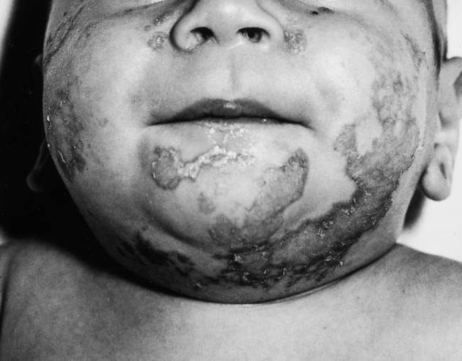
Physical examination includes assessment of growth, nutritional status and pubertal development. Plotting percentile charts is mandatory. A child who is growing normally is unlikely to be suffering from serious gastrointestinal disease. Plotting longitudinal measurements, if available, is very important as it may give clues to the onset of disease and could indicate the diagnosis. Other physical signs of malabsorption and specific nutritional deficiencies include: loss of muscle bulk and subcutaneous fat; peripheral oedema (hypoproteinaemia); bruising (vitamin K deficiency); glossitis and angular stomatitis (iron deficiency); finger clubbing (cystic fibrosis, Crohn’s disease, coeliac disease); skin rashes in coeliac disease (dermatitis herpetiformis) and inflammatory bowel disease (erythema nodosum, pyoderma gangrenosum); and specific skin disorders associated with zinc, vitamin A and essential fatty acid deficiencies (Fig. 20.3.1). Rickets is uncommon in our community, although biochemical vitamin D deficiency is quite common and is prevalent in patients with malabsorption, such as in cholestatic liver disease, and coeliac disease. It is important to examine carefully as there are many extraintestinal manifestations of gastrointestinal disease and malnutrition.


Stool examination


Stool examination is very simple and provides very important information. The presence of numerous white and red cells indicates colitis. This is usually due to bacterial or parasitic infection, to chronic inflammatory disorders of the large bowel, or to milk protein intolerance when identified in infants. Leukocytes are not increased in the stool of individuals with small bowel or pancreatic disease. Cysts of parasites such as Giardia lamblia indicate giardiasis.


Oil droplets seen on stool microscopy are always abnormal outside the newborn period and usually indicate fat maldigestion, as occurs with pancreatic insufficiency, for example in cystic fibrosis. Mucosal disease, such as coeliac disease, in general does not interfere with fat digestion because pancreatic function is usually normal. Mucosal disease interferes with the absorption of triglyceride products. These products are observed as fatty acid crystals on polarizing microscopy.


The presence of carbohydrate in the stool can be detected with Clinitest tablets. This is a commercially available bedside test in which the reaction between stool sugars such as lactose causes a colour change when added to the tablets. Greater than 500  mg/dL (0.5%) indicates carbohydrate malabsorption. Measurement of stool electrolytes and osmolality in the stool water is also a very useful test. When the sum of the stool electrolytes (i.e. sodium + potassium + chloride + bicarbonate) equals measured osmolality, a secretory diarrhoea is present. If the sum of the electrolytes is substantially less than the measured osmolality (>  100  mosmol/L), this indicates an osmotic diarrhoea.


Malabsorption with chronic diarrhoea


Diarrhoea is the most common presentation of malabsorption. Diarrhoea can be defined as increased frequency, fluidity and volume of stool. The following discussion will provide a systematic approach to the child with malabsorption and diarrhoea based on the type of stool, i.e.:


Some illustrative cases will be provided and a summary of this approach can be seen in Figure 20.3.2.


Fatty diarrhoea (steatorrhoea)


The differential diagnosis of fat malabsorption is quite wide ranging (see Box 20.3.1); however, if one understands the normal physiology of fat digestion and absorption, the differential diagnosis is much less daunting. Conditions that cause steatorrhoea can also be associated with protein maldigestion and/or malabsorption, although symptoms most commonly relate to the malabsorption of fat. The presence of fat in the stool is also more readily observed than protein.


image Clinical example


Mary was 9 months old. She presented with poor weight gain, chronic diarrhoea and a history of recurrent respiratory illnesses, including one admission at age 3 months with ‘bronchiolitis’. Loose stools were found each time her nappy was changed. On occasion her mother had noted oil drops in the stool. Despite the poor weight gain, Mary had an excellent appetite and was described as a voracious eater. She consumed a mixed diet, including infant formula, appropriate for age. Cereal was introduced at age 6 months. Mother also commented that she tasted salty when she kissed Mary.


On examination, Mary was found to be a thin wasted girl. Her height was on the 50th percentile and her weight was less than the 3rd percentile. She had mild finger clubbing, peripheral oedema, pallor of the tongue and palmar creases, but no signs of chronic liver disease. There was no abdominal distension of note, although she had a fine scaling rash over her trunk. Respiratory examination was normal. No other abnormal physical signs were present.


Results of investigations included haemoglobin 85  g/L (normal range 110–140) with a normocytic normochromic film, normal white cell count and differential; albumin 24  g/L (normal range 34–44) and normal liver function test results. Stool microscopy revealed copious fat droplets. Three-day faecal fat excretion estimation demonstrated an output of 35% of ingested fat (normal <  7% of intake).


Mary’s diarrhoea was due to fat malabsorption, as evidenced by her mother’s observation of fat droplets in the stool.


Mary’s sweat test demonstrated a sweat chloride of 80  mmol/L (a result >  60  mmol/L is diagnostic of cystic fibrosis). Genetic testing indicated that she was homozygous ΔF508 (the commonest mutation), consistent with her relatively severe symptoms. Introduction of pancreatic exocrine replacement therapy, a high-fat diet and vitamin supplements alleviated her diarrhoea and eventually corrected Mary’s failure to thrive, anaemia and skin rash.


Fat and protein digestion and absorption


Ingested fat in the form of triglycerides, cholesterol and phospholipids is, to a large extent, digested in the lumen of the small intestine and absorbed in the jejunum. This requires bile salts, which form micelles and solubilize the fat; pancreatic enzymes, such as lipase and co-lipase, which digest the fat; and an intact intestinal mucosa, which is required for absorption of the products of digestion. Following digestion in the micelles, breakdown products diffuse across the enterocyte apical membrane and are reconstituted in the cell into chylomicrons. These are small packets of triglyceride, phospholipid and cholesterol that associate with carrier proteins, such as β-lipoprotein, essential for cellular trafficking of the chylomicrons. After the chylomicrons are reconstituted, they exit the mucosa into the lymphatic system and subsequently pass into the systemic circulation. Some small-chain triglycerides can bypass this system and enter the portal venous system directly.


Protein digestion begins in the stomach by the action of pepsin and acid. However, most protein hydrolysis occurs in the lumen of the jejunum by the action of pancreatic proteases. These are secreted as inactive precursors. Chymotrypsin is converted to trypsin by the action of the small intestinal enzyme enterokinase. Activated trypsin further activates chymotrypsin and other proteases, such as carboxypeptidase. The products of protein hydrolysis are amino acids and oligopeptides. The latter are further hydrolysed to mono-, di- and tri-peptides by brush border hydrolases and are absorbed by specific membrane transporters. Di- and tri-peptides undergo hydrolysis to amino acids in the cytoplasm of the enterocyte. Isolated protein maldigestion/malabsorption is extremely rare. It usually occurs in association with malabsorption of other macronutrients.

Stay updated, free articles. Join our Telegram channel

Aug 4, 2016 | Posted by in PEDIATRICS | Comments Off on Chronic diarrhoea and malabsorption

Full access? Get Clinical Tree

Get Clinical Tree app for offline access