Chromosomal Abnormalities
George E. Tiller
For more than 50 years, chromosomal abnormalities have been recognized as the basis of certain genetic syndromes. Initially, only aneuploidies (the presence of more or fewer than 46 chromosomes) could be detected. Now, with the availability of fluorescence in situ hybridization (FISH) and DNA methylation testing, microdeletions and uniparental disomy (UPD) can be documented. The advent of microarray-based analysis (comparative genomic hybridization, or CGH) takes resolution to an even finer level, and introduces the confounding phenomenon of copy number variation. We have learned that although we carry two copies of each autosomal gene, the normal process of imprinting fine tunes the expression of certain regions of many chromosomes by silencing one copy of certain genes by DNA methylation. Consequently, UPD (presumably caused by trisomic “rescue” after fertilization) may result in the silencing of both copies of a gene or contiguous group of genes, despite the normal complement of 46 intact chromosomes. Further refinements in cytogenetic techniques, in tandem with molecular genetics, are making it possible to unravel normal processes in human development and pathologic processes in the origin of cancer.
The objectives of this chapter are to help the reader to:
Identify several indications of karyotyping.
Describe the clinical features of the more common chromosomal anomalies.
Appreciate the concepts of contiguous gene syndromes, imprinting, and UPD.
INCIDENCE OF CHROMOSOMAL DISORDERS
Chromosomal disorders are:
Found in >7% of human conceptuses
Responsible for >50% of miscarriages (trisomy 16 > 45, XO > others)
Found in 1 in 200 liveborn infants
INDICATIONS FOR CHROMOSOMAL ANALYSIS
Neonatal Period and Infancy (0-3 Years)
Phenotype of chromosomal anomaly (+21, +18, +13, XO)
Multiple congenital anomalies
Ambiguous genitalia
Certain tumors (retinoblastoma, Wilms tumor)
Childhood (4-10 Years)
Mental retardation (MR) and multiple congenital anomalies
Phenotype of chromosomal anomaly (XO, XXY, XYY)
Adolescence (11-20 Years)
Primary amenorrhea
Abnormal stature
Adults (20> Years)
Infertility with or without habitual abortion
Familial chromosomal aberration
Leukemia or certain tumors
Pregnancy at advanced maternal age
Pregnancy with risk for X-linked disorder
TYPES OF CHROMOSOMAL ABNORMALITIES
Numeric Changes
Of sets: triploidy, tetraploidy (all lethal)
Individual: trisomy, monosomy, sex chromosome aneuploidy
Mosaicism
Structural Changes
Deletions
Duplications
Translocations
Inversions
Functional Changes
Methylation defects (often caused by UPD)
EXAMPLES OF CHROMOSOMAL DISORDERS
Trisomy 21 (Down Syndrome)
Incidence: 1 in 750 (most common recognizable cause of MR)
Growth and development: May or may not be small-forgestational-age; short stature, MR
Central nervous system (CNS): Hypotonia, delayed motor skills, premature aging
Craniofacial: Flat face and occiput, upward-slanting palpebral fissures, epicanthal folds, Brushfield spots, prominent tongue, small ears, depressed nasal bridge
Extremities: Simian creases, clinodactyly, short fingers, increased space between the first and second toes, increased ulnar loops
Cardiac: Congenital heart disease in >50% of patients, including ventricular septal defect, endocardial cushion defects (atrioventricular canal)
Abdominal: Umbilical hernia, diastasis recti, duodenal obstruction
Skin/hair: Thin hair
Remarks: Increased risk of hypothyroidism, leukemia; all trisomies associated with advanced maternal age
Trisomy 13
Incidence: 1 in 5000
Growth and development: Small-for-gestational-age, intrauterine growth retardation, failure to thrive, severe MR
CNS: Hypotonia, seizures, apnea, deafness, holoprosencephaly
Craniofacial: Microcephaly, microphthalmia, colobomata, cleft lip and palate, abnormal ears
Extremities: Polydactyly (Fig. 41.1), flexion deformities, clubfoot
Cardiac: Ventricular septal defect, patent ductus arteriosus, atrial septal defect, coarctation of the aorta
Abdominal: Umbilical hernia, omphalocele, single umbilical artery
Renal: Polycystic kidneys
Skin/hair: Occipital scalp defects (cutis aplasia), hemangiomas
Remarks: Survival rate beyond the first year is <20%
Trisomy 18
Incidence: 1 in 3000
Growth and development: Small-for-gestational-age, intrauterine growth retardation, failure to thrive, severe MR
CNS: Hypertonia
Craniofacial: Prominent occiput; low-set, malformed ears; micrognathia; cleft lip and palate (Fig. 41.2)
Extremities: Overlapping fingers, rocker-bottom feet, clubfoot (Fig. 41.3)
Cardiac: Ventricular septal defect, patent ductus arteriosus, atrial septal defect
Abdominal: Inguinal, umbilical hernias
Renal: Various anomalies
Skin/hair: Single flexion crease on digits
Remarks: Survival rate beyond the first year is <20%
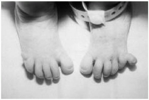 Figure 41.1 Newborn with trisomy 13. Note the postaxial polydactyly on left foot.
Stay updated, free articles. Join our Telegram channel
Full access? Get Clinical Tree
 Get Clinical Tree app for offline access

|